

Health Dictionary
Reliable, easy-to-read patient information on common medical conditions — written in plain language, free of jargon, and grounded in current evidence. Browse the most frequent diagnoses, understand what they mean for everyday life, and learn which steps and questions help you have a focused conversation with your doctor.
Mastitis other imaging findings
Introduction
Overview
Overview
On mammography, bacterial mastitis usually appears as ill-defined regions of increased density and skin thickening.[1][2][3]
Other Imaging Findings of Mastitis
Other Imaging Findings of Mastitis
On mammography, bacterial (puerperal or non-puerperal) mastitis usually shows ill-defined regions of increased density and skin thickening[1][2][3] as shown below:.
-
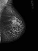 Mammogram of Mastitis – Case courtesy of Dr Maxime St-Amant, Radiopaedia.org, rID: 26843

References
References
- ↑ 1.0 1.1 Gautier N, Lalonde L, Tran-Thanh D, El Khoury M, David J, Labelle M; et al. (2013). “Chronic granulomatous mastitis: Imaging, pathology and management”. Eur J Radiol. 82 (4): e165–75. doi:10.1016/j.ejrad.2012.11.010. PMID 23200627.
- ↑ 2.0 2.1 Tan H, Li R, Peng W, Liu H, Gu Y, Shen X (2013). “Radiological and clinical features of adult non-puerperal mastitis”. Br J Radiol. 86 (1024): 20120657. doi:10.1259/bjr.20120657. PMC 3635790. PMID 23392197.
- ↑ 3.0 3.1 Jari I, Naum AG, Ursaru M, Manafu EG, Gheorghe L, Negru D (2015). “BREAST INFECTIONS: DIAGNOSIS WITH ULTRASOUND AND MAMMOGRAPHY”. Rev Med Chir Soc Med Nat Iasi. 119 (2): 419–24. PMID 26204646.
Editor-In-Chief: C. Michael Gibson, M.S., M.D. [1]; Associate Editor(s)-in-Chief: Prince Tano Djan, BSc, MBChB [2]