

Health Dictionary
Reliable, easy-to-read patient information on common medical conditions — written in plain language, free of jargon, and grounded in current evidence. Browse the most frequent diagnoses, understand what they mean for everyday life, and learn which steps and questions help you have a focused conversation with your doctor.
Mature cystic teratoma
Introduction
Overview
Overview
Mature cystic teratomas are cystic tumors composed of well-differentiated derivations from at least two of the three germ cell layers (ectoderm, mesoderm, and endoderm). They affect a younger age group (mean patient age, 30 years) than epithelial ovarian neoplasms. Mature cystic teratoma is the most common germ cell neoplasm and, in some series, the most common ovarian neoplasm. The tumors are bilateral in about 10% of cases. Most mature cystic teratomas are asymptomatic. Complications include rupture, malignant degeneration, or (most commonly) ovarian torsion.
Imaging Findings
Imaging Findings
- Most mature cystic teratomas can be diagnosed at US.
- The most common manifestation is a cystic lesion with a densely echogenic tubercle (Rokitansky nodule) projecting into the cyst lumen.
- The second manifestation is a diffusely or partially echogenic mass with the echogenic area usually demonstrating sound attenuation owing to sebaceous material and hair within the cyst cavity.
- The third manifestation consists of multiple thin, echogenic bands caused by hair in the cyst cavity.
- The diagnosis of mature cystic teratoma at CT and MR imaging is fairly straightforward because these modalities are more sensitive for fat than US.
Patient #1: US and MR images demonstrate a large dermoid
-

-

-

-

-
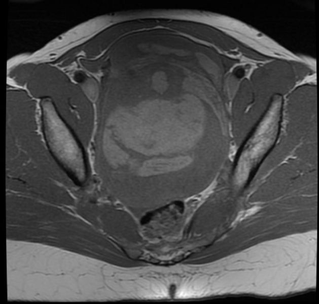 T1
T1 -
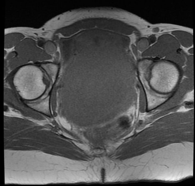 T1
T1 -
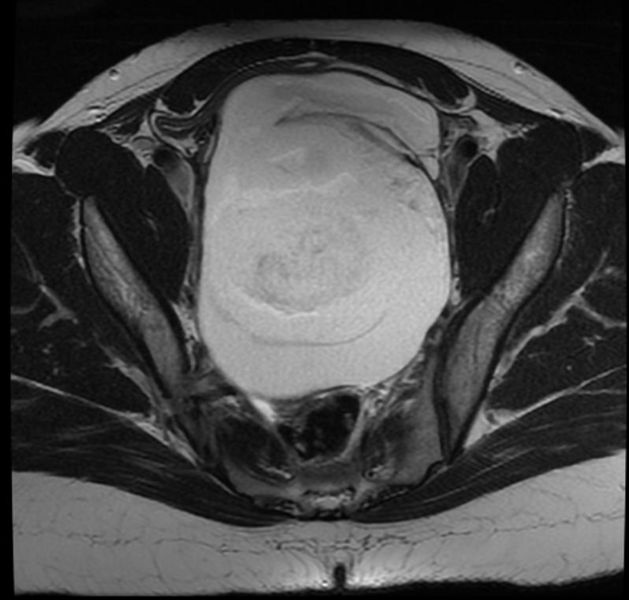 T2
T2 -
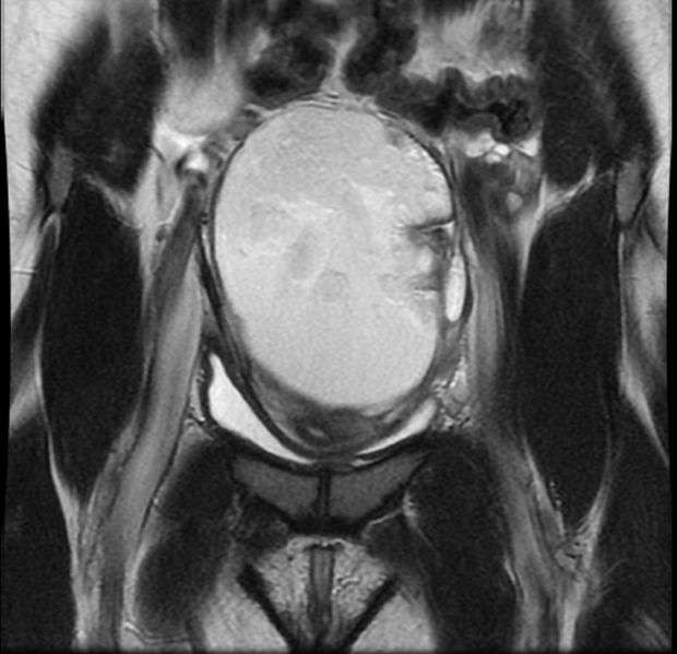 T2
T2 -
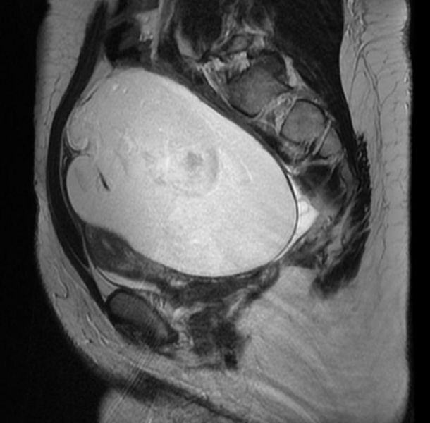 T2
T2 -
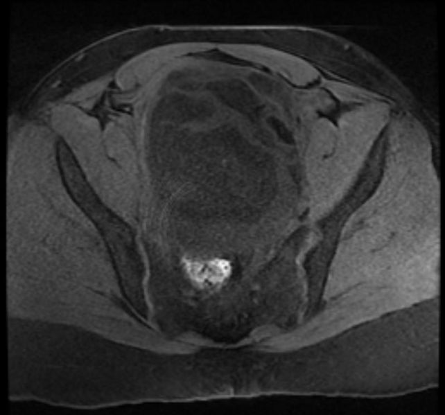 T1 fat sat
 -
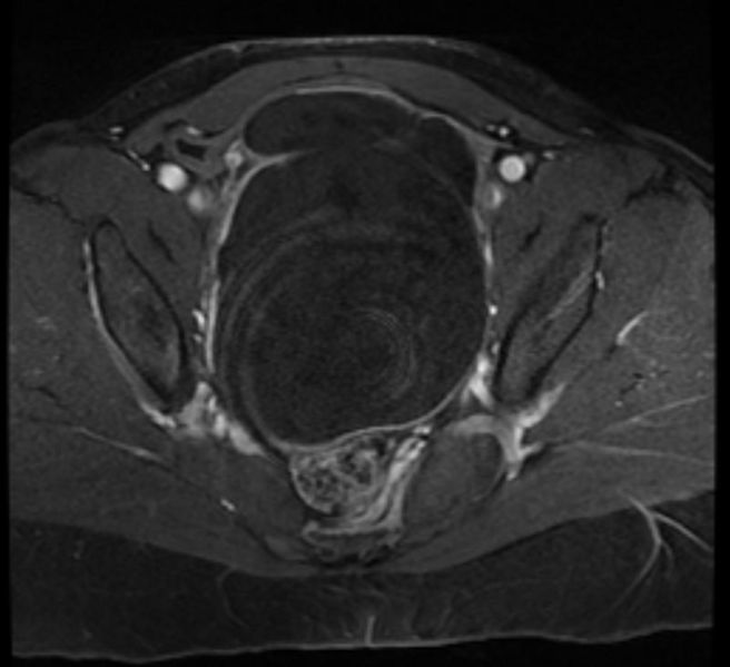 T1 fat sat post contrast
 -
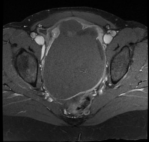 T1 fat sat post contrast


Patient #2: CT images demonstrate left ovarian mature cystic teratoma


References
References
Editor-In-Chief: C. Michael Gibson, M.S., M.D. [1]