
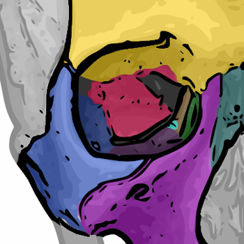
Maxilla
Editor-In-Chief: C. Michael Gibson, M.S., M.D. [1]
Overview
Overview
The maxilla (plural: maxillae) is a fusion of two bones along the palatal fissure that form the upper jaw. This is similar to the mandible, which is also a fusion of two halves at the mental symphysis.
Function
Function
The alveolar process of the maxilla holds the upper teeth, and is referred to as the maxillary arch. The maxilla attaches laterally to the zygomatic bones (cheek bones).
The maxilla assists in forming the boundaries of three cavities:
- the roof of the mouth
- the floor and lateral wall of the nasal antrum
- the floor of the orbit
The maxilla also enters into the formation of two fossae: the infratemporal and pterygopalatine, and two fissures, the inferior orbital and pterygomaxillary.
Components
Components
Each half of the fused maxilla consists of:
- The body of the maxilla
- Four processes
- The zygomatic process
- The frontal process
- The alveolar process
- The palatine process
- Infraorbital foramen
Articulations
Articulations
The maxilla articulates with nine bones:
- two of the cranium: the frontal and ethmoid
- seven of the face: the nasal, zygomatic, lacrimal, inferior nasal concha, palatine, vomer, and the adjacent fused maxillary bone.
Sometimes it articulates with the orbital surface, and sometimes with the lateral pterygoid plate of the sphenoid.
Additional images
Additional images
-
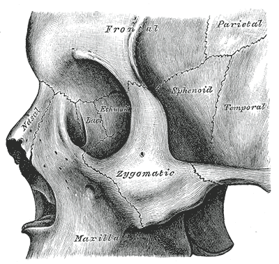
 The seven bones which articulate to form the orbit.
 -
 Facial bones.
 -
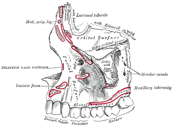 Left maxilla. Outer surface.
 -
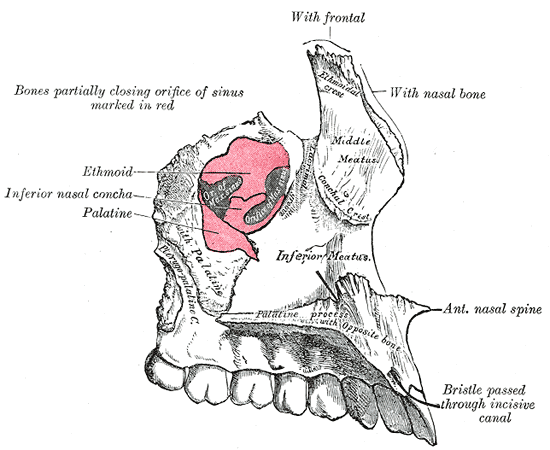 Left maxilla. Nasal surface.
 -
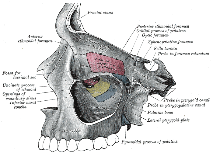 Left maxillary sinus opened from the exterior.
 -
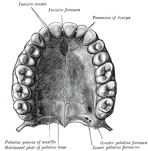 The bony palate and alveolar arch.
 -
 Sphenoid bone visible center right.
 -
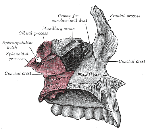 Articulation of left palatine bone with maxilla.
 -
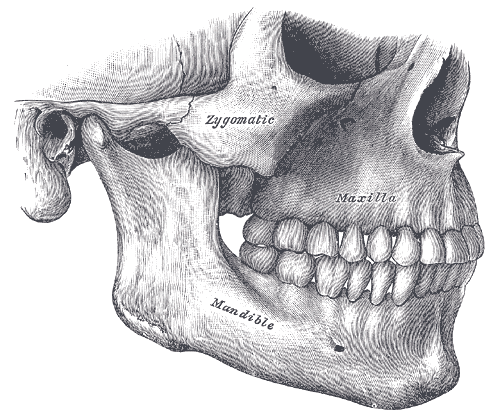 Side view of the teeth and jaws.


See also
See also
References
References
Looking for the patient version?
© 2026 MyEClinic – IFTM Institut für Telematik in der Medizin GmbH