
Melorheostosis
Editor-In-Chief: C. Michael Gibson, M.S., M.D. [1]
Overview
Overview
Melorheostosis is usually discovered in childhood, occasionally in adulthood. The male-to-female ratio is usually 1:1. It can occur in a single limb, and the lower extremity is more commonly affected than the upper extremity. The epicenter is periosteal or endosteal. The appearance consists of cortical hyperostosis in one or multiple bones, often with intervening soft-tissue calcification or ossification. The limb involved with melorheostosis often demonstrates joint pain, swelling, and limitation of motion in childhood. There is often associated growth disturbance, muscular contraction, and limb length discrepancy. There may be overlying skin changes. At pathologic analysis, thickened and enlarged osseous trabeculae are noted, associated with fibrous tissue replacement of the marrow space. There is controversy involving the distribution of melorheostosis. It has been suggested that the distribution mimics that of the sclerotomes (zones supplied by individual spinal sensory nerves), implying a neurogenic origin.
Diagnosis
Diagnosis
The imaging findings are
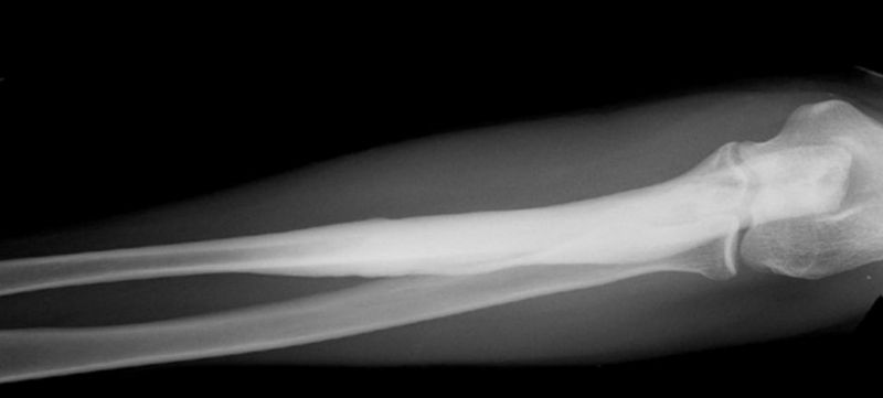
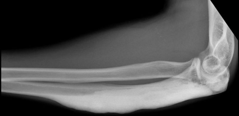
- At radiography, contiguous bones of an extremity are often involved, although there may be involvement of a single bone.
- There is cortical hyperostosis with intervening soft-tissue calcification or ossification. Dripping candle wax appearance.
- There may be endosteal hyperostosis with obliteration of the medullary space.
-
-
-
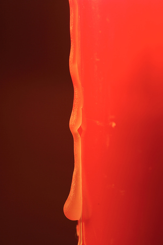 Dripping candle wax


Looking for the patient version?
© 2026 MyEClinic – IFTM Institut für Telematik in der Medizin GmbH