
Mitral regurgitation chest x-ray
Editor-In-Chief: C. Michael Gibson, M.S., M.D. [1]; Associate Editor(s)-in-Chief: Cafer Zorkun, M.D., Ph.D. [2]; Varun Kumar, M.B.B.S.; Lakshmi Gopalakrishnan, M.B.B.S.
Overview
Overview
The chest X-ray in individuals with chronic mitral regurgitation (MR) is characterized by the presence of an enlargement of the left atrium and the left ventricle. In acute MR, pulmonary edema is present and the heart is not enlarged.
Chest X Ray
Chest X Ray
- In acute MR, chest X-ray reveals pulmonary edema caused by pulmonary congestion secondary to the abrupt rise in pressure in the left atrium. There is no enlargement of the cardiac silhouette in acute MR.[1]
- In chronic MR, the pulmonary vascular markings are typically normal since the pulmonary venous pressures are usually not significantly elevated. The left side of the heart might be enlarged. The left main bronchus can be pushed up by an enlarged left atrium.
- Calcification of the mitral annulus is not infrequent, and is seen in up to 35% of elderly patients on echocardiography which is more sensitive than chest X-ray. It typically begins around the margins of the posterior leaflet forming a “C”, eventually the “C” closes forming an “O” with anterior leaflet involvement. [3])
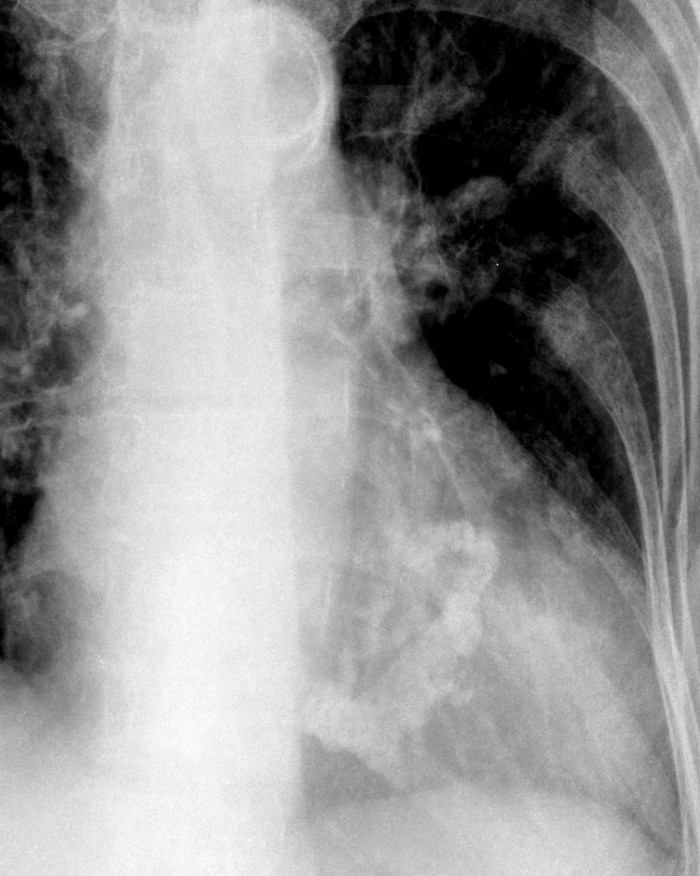
Calcification of the mitral annulus

References
References
- ↑ Mitral regurgitation https://radiopaedia.org/articles/mitral-valve-regurgitation (2016) Accessed on December 13, 2016
Looking for the patient version?
© 2026 MyEClinic – IFTM Institut für Telematik in der Medizin GmbH