
Osteomyelitis x ray
Editor-In-Chief: C. Michael Gibson, M.S., M.D. [1]; Associate Editor(s)-in-Chief: Seyedmahdi Pahlavani, M.D. [2]
Overview
Overview
The diagnosis of osteomyelitis is often based on radiologic results which demonstrate a lytic center with a ring of sclerosis, though bone cultures are normally required to identify the specific pathogen. Conventional radiographic evaluation of acute osteomyelitis is insufficient because bone changes are not evident for 14–21 days after the onset of infection.
X Ray
X Ray
- Conventional radiography is readily available, relatively inexpensive, and useful in the differentiation of infection from trauma and tumors, and it is the initial imaging test of choice for suspected osteomyelitis.
- In addition, plain radiography is often a helpful adjunct to secondary imaging studies. Unfortunately, radiographic evidence of osteomyelitis lags behind the clinical picture, and less than one third of patients have abnormalities on plain radiographs in the first 7 to 10 days after the onset of symptoms.
- Radiologic bone findings suggestive of osteomyelitis is a lytic center with a ring of sclerosis.[1]
- Other findings include soft tissue edema and deep muscle displacement.
-
-
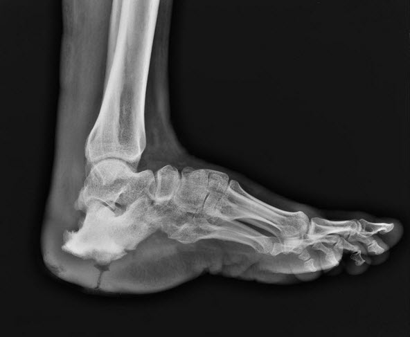 Air filled sinus tract which leads to to sclerosed, deformed calcaneum.
 -
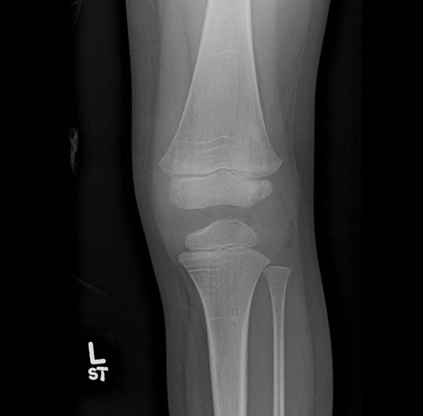
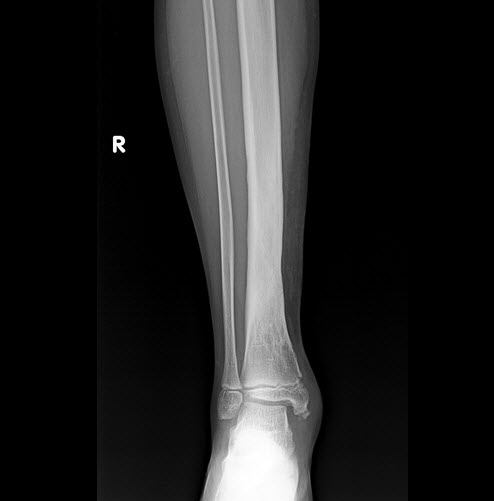
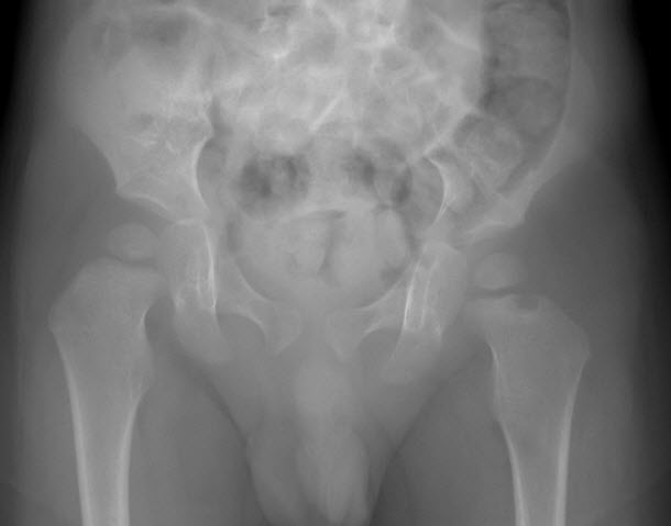 Lucency on the lateral margin of the metaphysis adjacent to the physis of head of left femor.
 -
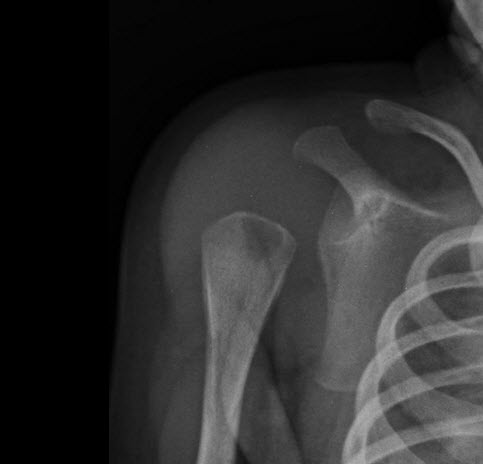 Proximal humeral metaphyseal lytic focus in a 25 days neonate.
 -
-
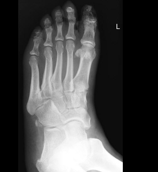 Loss of soft tissue over the great toe with lucency in the surrounding soft tissue, associated patchy osteoporosis in the underlying phalanx in a patient with diabetic foot.


References
References
- ↑ Pineda C, Vargas A, Rodríguez AV (2006). “Imaging of osteomyelitis: current concepts”. Infect. Dis. Clin. North Am. 20 (4): 789–825. doi:10.1016/j.idc.2006.09.009. PMID 17118291.
Looking for the patient version?
© 2026 MyEClinic – IFTM Institut für Telematik in der Medizin GmbH