

Health Dictionary
Reliable, easy-to-read patient information on common medical conditions — written in plain language, free of jargon, and grounded in current evidence. Browse the most frequent diagnoses, understand what they mean for everyday life, and learn which steps and questions help you have a focused conversation with your doctor.
Osteosarcoma case study one
Introduction
Overview
Overview
Case Studies
Case Studies
Case #1
Clinical Summary
This 14-year-old white male first experienced mild pain in the left knee after playing baseball, approximately two months prior to admission. The pain persisted in an intermittent fashion, and was described as being somewhat worse at night. Approximately two weeks prior to admission, the pain increased significantly and was accompanied by marked swelling and loss of considerable motion of the knee joint. These symptoms were accompanied by a history of decreased appetite, lethargy, and a 10-pound weight loss. On physical examination, the left knee was enlarged diffusely, firm, and non-tender. Following biopsy, the patient was subjected to surgical removal of the distal femur and knee with placement of a prosthetic knee joint and bone grafts.
Autopsy Findings
The distal diaphysis of the femur and adjacent soft tissues were involved in a 15 x 10 x 10-cm mass. The cut surface of the mass was fleshy white, with focal areas of hemorrhage.
Pathological Findings
-
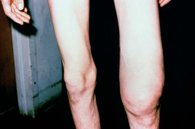 This is a photograph of the patient prior to surgery. Note the marked swelling of the knee.
 -
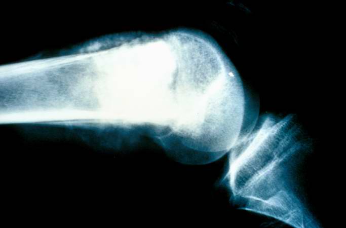 This is a radiograph showing the tumor in the distal femur.
 -
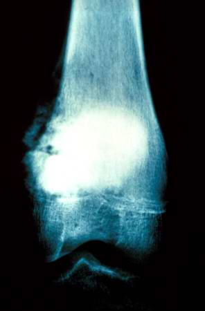 This is another view of the tumor in the distal femur.
 -
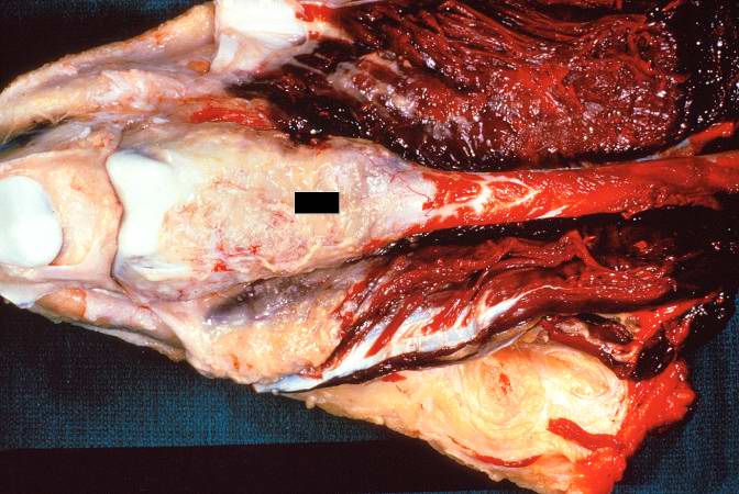 This is a gross photograph of the surgical specimen with tissue dissected away to demonstrate the tissue mass.
 -
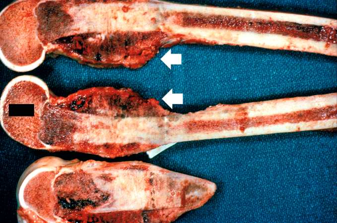 These are cut sections of the distal femur containing the tumor. The periosteal involvement is evident from this picture (arrows).
 -
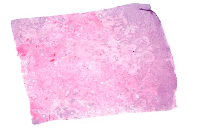 This is a low-power photomicrograph of decalcified histologic section from this tumor. Note the blue color (cell nuclei stain blue) of much of this section indicating the increased cellularity of the tumor.
 -
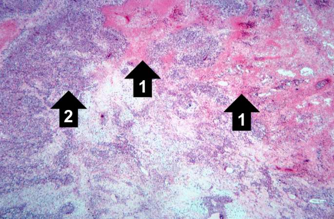 This is a higher-power photomicrograph of decalcified histologic section from this tumor. There are areas of osteoid (1) and cellular areas (2).
 -
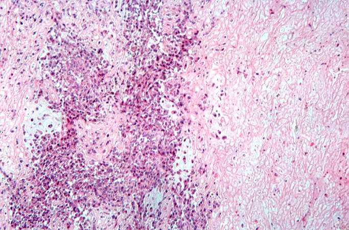 This is a high-power photomicrograph of decalcified histologic section showing the cellularity of the tumor.


References
References
Editor-In-Chief: C. Michael Gibson, M.S., M.D. [1]