
Ovarian fibroma
Editor-In-Chief: C. Michael Gibson, M.S., M.D. [1]
Contributors: Cafer Zorkun M.D., PhD.
Overview
Overview
Ovarian fibromas account for approximately 4% of all ovarian neoplasms and are the most common sex cord tumor. Fibromas are generally asymptomatic and typically detected in middle-aged women at palpation during routine gynecologic examination. They are associated with ascites in 40% of cases and with pleural effusions in a small percentage of cases. Meig syndrome consists of an ovarian fibroma with ascites and a pleural effusion. [1] [2]
Epidemiology
Epidemiology
Fibromas are seen in 75% of patients with nevoid basal cell carcinoma syndrome.
Diagnosis
Diagnosis
Ultrasonography
Fibromas most commonly manifest as solid, hypoechoic masses with sound attenuation; however, the US appearance is variable.
Computed Tomography
Fibromas manifest as diffuse, slightly hypoattenuating masses with poor, very slow contrast enhancement.
Magnetic Resonance Imaging
Fibromas demonstrate homogeneous, relatively low signal intensity on T1-weighted MR images. On T2-weighted images, fibromas appear as well-circumscribed masses with low signal intensity containing scattered high-signal-intensity areas representing edema or cystic degeneration.
Diagnostic Findings
Diagnostic Findings
Ultrasonography
-
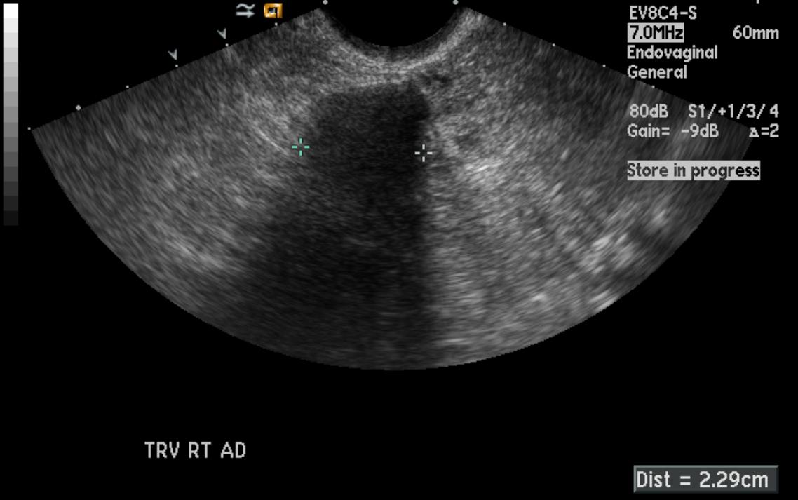 US: Ovarian fibroma.
 -
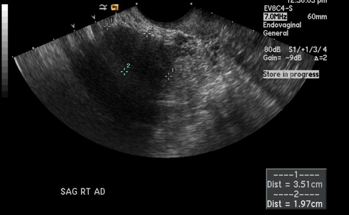 US: Ovarian fibroma.


Magnetic Resonance Imaging
-
 MRI: Ovarian fibroma.
 -
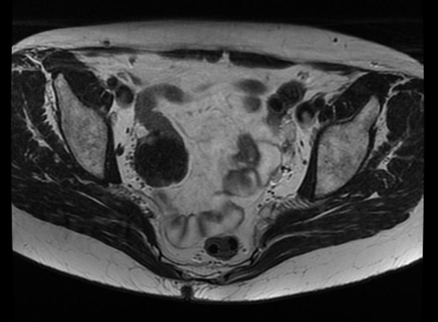 MRI: Ovarian fibroma.
 -
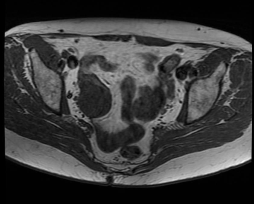 MRI: Ovarian fibroma.
 -
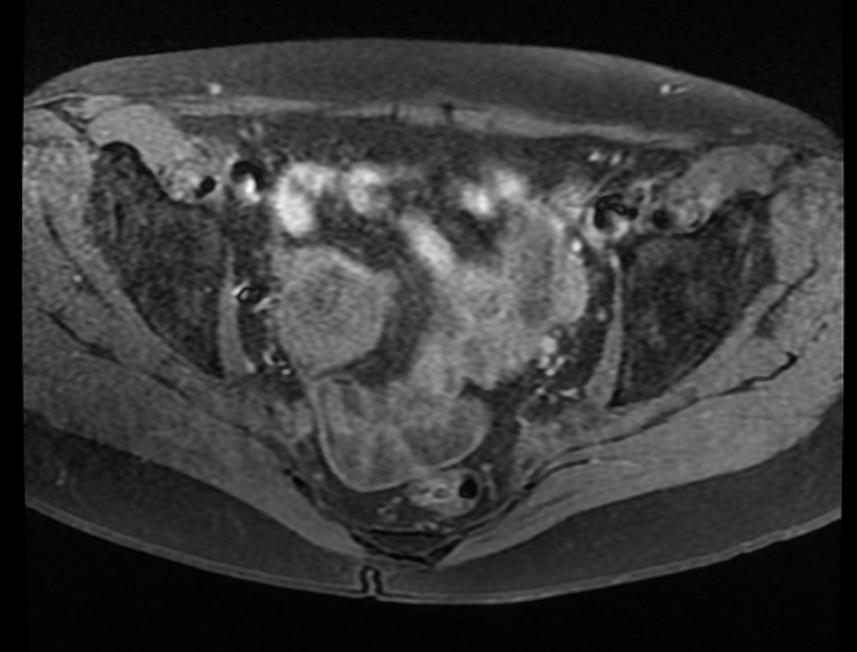 MRI: Ovarian fibroma.
 -
 MRI: Ovarian fibroma.


Pathology
-
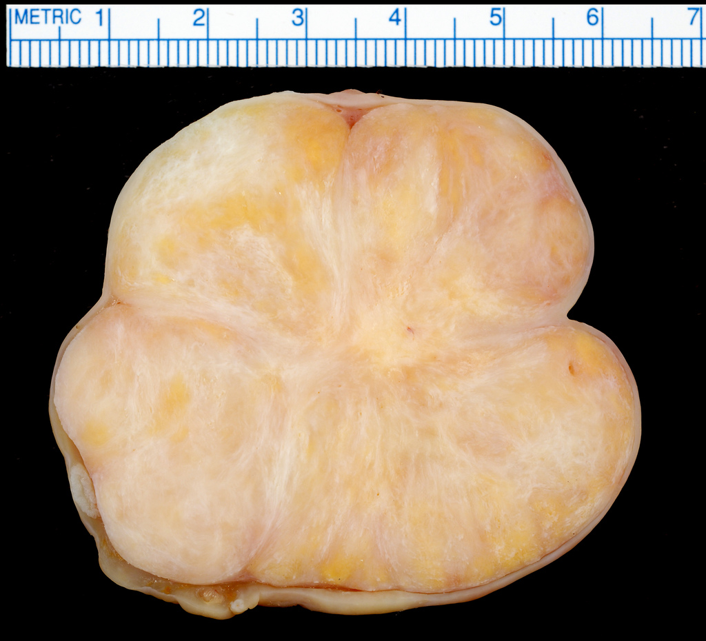 Ovarian Fibroma

References
References
- ↑ Ricardo B. Fonseca, and Ewa F. Grzeszczak. Case 128: Bilateral Ovarian Fibromas in Nevoid Basal Cell Carcinoma Syndrome. Radiology 2008 246: 318-321.
- ↑ Yong-Yeon Jeong, Eric K. Outwater, and Heoun Keun Kang. Imaging Evaluation of Ovarian Masses. RadioGraphics 2000 20: 1445-1470.
External Links
External Links
Looking for the patient version?
© 2026 MyEClinic – IFTM Institut für Telematik in der Medizin GmbH