

Health Dictionary
Reliable, easy-to-read patient information on common medical conditions — written in plain language, free of jargon, and grounded in current evidence. Browse the most frequent diagnoses, understand what they mean for everyday life, and learn which steps and questions help you have a focused conversation with your doctor.
Pancoast tumor echocardiography or ultrasound
Introduction
Overveiw
Overveiw
Pancoast tumor, a subtype of lung cancer located at the lung apex. On endobronchial and endoscopic ultrasound, characteristic findings of lung cancer may include: enlarged lymph nodes and local invasion to adjacent bronchial structures and mediastinum. Endobronchial ultrasound is a first-line diagnostic modality for mediastinal staging.[1]
Ultrasound
Ultrasound
- Endobronchial ultrasound is a first-line diagnostic modality for mediastinal staging.[1]
- Common features of endobronchial ultrasound, include:[2]
- Evaluation of lymph nodes and other structures in the mediastinum
- Mediastinum invasion staging
- Determination of management strategy
- Real time evaluation of structures
- Sensitivity 90% and specificity of 97%
- On ultrasound (endobronchial ultrasound), findings of Pancoast tumor may include:[3]
- Enlarged lymph nodes
- Local invasion to adjacent bronchial structures and mediastinum
- The table below summarizes the advantages and disadvantages of both ultrasound modalities, for the diagnostic assessment of Pancoast tumor.
Procedure Advantages Disadvantages Endobronchial ultrasound (EBUS) - Direct visualization of lymph node stations
- Complements endoscopic ultrasound: covers lymph node stations 2R and 4R which are difficult to access by endoscopic ultrasound
- Lower false-negative rate than with blind transbronchial fine needle aspiration and fewer complications
- More invasive than endoscopic ultrasound, few practitioners, but rapidly growing in popularity
Endoscopic ultrasound (EUS) - Least invasive modality
- Uses the esophagus to access mediastinal lymph nodes
- Excellent for staging lymph nodes
- Useful for station 2L and 4L, L adrenal, celiac lymph node
- Cannot reliably access right sided paratracheal lymph node stations 2 R and 4R
- Accurate discrimination of primary hilar tumors and involved lymph nodes is important
-
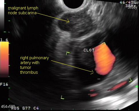 Doppler endoscopic ultrasound: mediastinal lymph node
 -
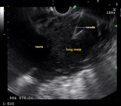 Endoscopic ultrasound: A biopsy window is found and an fine needle aspiration advanced into the mass
 -
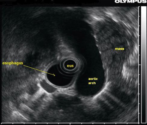 Endoscopic ultrasound: A lung mass which is partially behind the aorta is seen with endoscopic ultrasound


References
References
- ↑ 1.0 1.1 Kinsey CM, Arenberg DA (2014). “Endobronchial ultrasound-guided transbronchial needle aspiration for non-small cell lung cancer staging”. Am. J. Respir. Crit. Care Med. 189 (6): 640–9. doi:10.1164/rccm.201311-2007CI. PMID 24484269.
- ↑ Tests for non-small cell lung cancer. American Cancer Society. http://www.cancer.org/cancer/lungcancer-non-smallcell/detailedguide/non-small-cell-lung-cancer-diagnosis Accessed on February 25, 2016
- ↑ Lung cancer staging. Wikipedia. https://en.wikipedia.org/wiki/Lung_cancer_staging Accessed on February 25,2016
Editor-In-Chief: C. Michael Gibson, M.S., M.D. [1]; Associate Editor(s)-in-Chief: Mazia Fatima, MBBS [2]