

Health Dictionary
Reliable, easy-to-read patient information on common medical conditions — written in plain language, free of jargon, and grounded in current evidence. Browse the most frequent diagnoses, understand what they mean for everyday life, and learn which steps and questions help you have a focused conversation with your doctor.
Patellar ligament
Introduction
Overview
Overview
The Patellar ligament (anterior ligament) is the central portion of the common tendon of the Quadriceps femoris, which is continued from the patella to the tuberosity of the tibia.
It is a strong, flat, ligamentous band, about 8 cm. in length, attached, above, to the apex and adjoining margins of the patella and the rough depression on its posterior surface; below, to the tuberosity of the tibia; its superficial fibers are continuous over the front of the patella with those of the tendon of the Quadriceps femoris.
The medial and lateral portions of the tendon of the Quadriceps pass down on either side of the patella, to be inserted into the upper extremity of the tibia on either side of the tuberosity; these portions merge into the capsule, as stated above, forming the medial and lateral patellar retinacula.
The posterior surface of the ligamentum patellæ is separated from the synovial membrane of the joint by a large infrapatellar pad of fat, and from the tibia by a bursa.
Additional images
Additional images
-
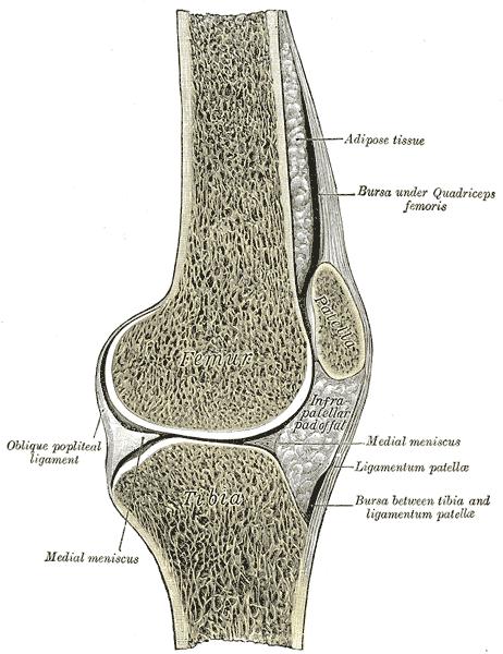 Sagittal section of right knee-joint.
 -
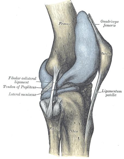 Capsule of right knee-joint (distended). Lateral aspect.


External links
External links
- Template:SUNYAnatomyFigs – “Muscles of the anterior (extensor) compartment of the leg.”
- Template:EMedicineDictionary