

Health Dictionary
Reliable, easy-to-read patient information on common medical conditions — written in plain language, free of jargon, and grounded in current evidence. Browse the most frequent diagnoses, understand what they mean for everyday life, and learn which steps and questions help you have a focused conversation with your doctor.
Pneumoconiosis chest x ray
Introduction
Overview
Overview
An x-ray may be helpful in the diagnosis of pneumoconiosis. It is the initial screening test used however CT scan provides more sensitivity and specificity. [1] [2] [3] [4]
X Ray
X Ray
An x-ray may be helpful in the diagnosis of pneumoconiosis. Findings on an x-ray suggestive of/diagnostic of pneumoconiosis include:[5] [6]
- Pleural plaques are pathognomonic for asbestosis.
- Interstitial nodules in the upper zone of the lungs for both silicosis and coal worker’s pneumoconiosis.
- Progressive massive fibrosis is seen in both silicosis and coal worker’s pneumoconiosis.
- Hilar adenopathy with ground glass opacities is seen in berylliosis.
-
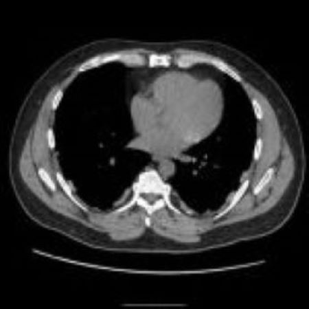 Asbestosis with pleural plaques – Case courtesy of Dr Hani Makky Al Salam, <a href=”https://radiopaedia.org/“>Radiopaedia.org</a>. From the case <a href=”https://radiopaedia.org/cases/45002“>rID: 45002</a>
 -
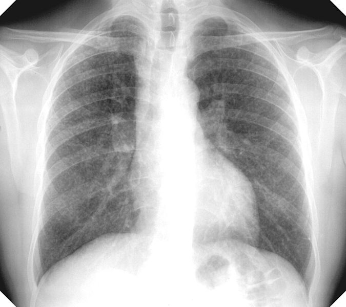 Silicosis with nodules
 -
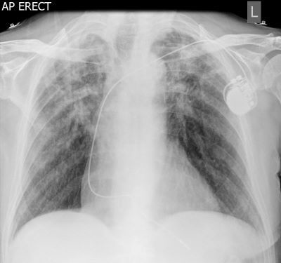 Pneumoconiosis with massive pulmonary fibrosis – Case courtesy of Dr Ian Bickle, <a href=”https://radiopaedia.org/“>Radiopaedia.org</a>. From the case <a href=”https://radiopaedia.org/cases/50303“>rID: 50303</a>


References
References
- ↑ Remy-Jardin M, Remy J, Farre I, Marquette CH (1992). “Computed tomographic evaluation of silicosis and coal workers’ pneumoconiosis”. Radiol Clin North Am. 30 (6): 1155–76. PMID 1410306.
- ↑ Akira M, Yokoyama K, Yamamoto S, Higashihara T, Morinaga K, Kita N; et al. (1991). “Early asbestosis: evaluation with high-resolution CT”. Radiology. 178 (2): 409–16. doi:10.1148/radiology.178.2.1987601. PMID 1987601.
- ↑ Copley SJ, Wells AU, Sivakumaran P, Rubens MB, Lee YC, Desai SR; et al. (2003). “Asbestosis and idiopathic pulmonary fibrosis: comparison of thin-section CT features”. Radiology. 229 (3): 731–6. doi:10.1148/radiol.2293020668. PMID 14576443.
- ↑ Walkoff L, Hobbs S (2020). “Chest Imaging in the Diagnosis of Occupational Lung Diseases”. Clin Chest Med. 41 (4): 581–603. doi:10.1016/j.ccm.2020.08.007. PMID 33153681 Check
|pmid=value (help). - ↑ “StatPearls”. 2021. PMID 32310362 Check
|pmid=value (help). - ↑ Wade WA, Petsonk EL, Young B, Mogri I (2011). “Severe occupational pneumoconiosis among West Virginian coal miners: one hundred thirty-eight cases of progressive massive fibrosis compensated between 2000 and 2009”. Chest. 139 (6): 1458–1462. doi:10.1378/chest.10-1326. PMID 20884728.
Editor-In-Chief: C. Michael Gibson, M.S., M.D. [1]; Associate Editor(s)-in-Chief: Dushka Riaz, MD