

Health Dictionary
Reliable, easy-to-read patient information on common medical conditions — written in plain language, free of jargon, and grounded in current evidence. Browse the most frequent diagnoses, understand what they mean for everyday life, and learn which steps and questions help you have a focused conversation with your doctor.
Pneumomediastinum CT
Introduction
Overview
Overview
The diagnosis can be confirmed via chest X-ray showing a radiolucent outline around the heart and mediastinum or via CT scanning of the thorax.
CT
CT
The following findings are associated with pneumomediastinum on CT-scan of the chest:[1][2][3][4][5][6][7]
- Subcutaneous emphysema
- Naclerio V sign
- Seen in pneumomediastinum occurring often secondary to an oesophageal rupture but it is not entirely specific to that condition.
- Pneumopericardium
- Gas anterior to pericardium
- Ring around artery sign
- Gas around pulmonary artery and main branches
- Tubular artery sign
- Gas outlining major aortic branches
- Double bronchial wall sign
- Gas outlining bronchial wall
- Continuous diaphragm sign
- Gas trapped posterior to pericardium
- Extrapleural sign
- Gas between parietal pleura and diaphragm
- Gas in pulmonary ligament
Pediatric pneumomediastinum has different appearances:
- Thymic wing sign
- Elevated thymus
- Haystack sign (the heart appears like a haystack in a Monet painting)
- Gas crossing the superior mediastinum
-
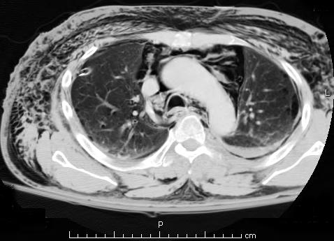 A CT scan showing air in the mediastinum

Images shown below are courtesy of RadsWiki.
-
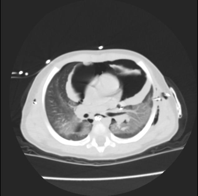 Pneumomediastinum
 -
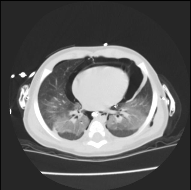 Pneumomediastinum


-
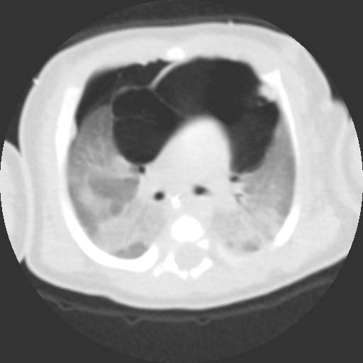 Pneumomediastinum and pneumothorax
 -
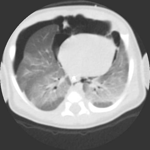 Pneumomediastinum and pneumothorax


-
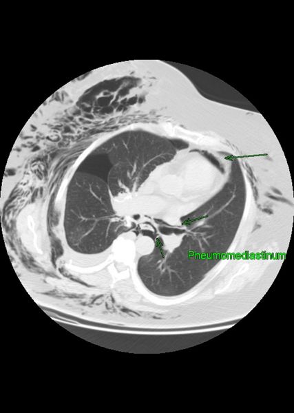 Pneumomediastinum and pneumothorax and subcutaneous emphysema

References
References
- ↑ Taveras, J., & Ferrucci, J. (1986). Radiology. Diagnosis/imaging/intervention. 5 volumes. Annual revision service. R. Health Professions,Philadelphia, PA.
- ↑ Sandler, Carl M.; Llbshltz, Herman I.; Marks, Gerald; Libshitz, Herman I. (1975). “Pneumoperitoneum, Pneumomediastinum and Pneumopericardium Following Dental Extraction”. Radiology. 115 (3): 539–540. doi:10.1148/15.3.539. ISSN 0033-8419.
- ↑ Kim, Hye Rin; Yoo, Seung Min; Lee, Hwa Yeon; Han, Jin Hee; Frazier, Aletta A; White, Charles S (2016). “Presence of subpleural pulmonary interstitial emphysema as an indication of single or multiple alveolar ruptures on CT in patients with spontaneous pneumomediastinum”. Acta Radiologica. 57 (12): 1483–1489. doi:10.1177/0284185116629830. ISSN 0284-1851.
- ↑ Moseley, John E. (1960). “Loculated Pneumomediastinum in the Newborn”. Radiology. 75 (5): 788–790. doi:10.1148/75.5.788. ISSN 0033-8419.
- ↑ Hammond DI (March 1984). “The “ring-around-the-artery” sign in pneumomediastinum”. J Can Assoc Radiol. 35 (1): 88–9. PMID 6725378.
- ↑ Levin, Bertram (1973). “The continuous diaphragm sign”. Clinical Radiology. 24 (3): 337–338. doi:10.1016/S0009-9260(73)80050-9. ISSN 0009-9260.
- ↑ Lillard, Richard L.; Allen, Parker (1965). “The Extrapleural Air Sign in Pneumomediastinum”. Radiology. 85 (6): 1093–1098. doi:10.1148/85.6.1093. ISSN 0033-8419.
Editor-In-Chief: C. Michael Gibson, M.S., M.D. [1] Associate Editor(s)-in-Chief: Trusha Tank, M.D.[2]