
Pneumoperitoneum
Editor-In-Chief: C. Michael Gibson, M.S., M.D. [1]
Overview
Overview
Pneumoperitoneum is air or gas in the abdominal (peritoneal) cavity[1], often seen on x-ray, but small amounts are often missed and CT is nowadays regarded as a criterion standard in the assessment of a pneumoperitoneum.[2].
Causes
Causes
The most common cause is a perforated abdominal viscus, generally a perforated ulcer, although any part of the bowel may perforate from a benign ulcer, tumor or trauma. A perforated appendix seldom causes a pneumoperitoneum. A pneumoperitoneum is deliberately created by the surgical team in order to perform laparoscopic surgery. This is achieved by insufflating the abdomen with carbon dioxide.
- Perforated peptic ulcer
- Bowel obstruction
- Ruptured diverticulum
- Penetrating trauma
- Ruptured inflammatory bowel disease (e.g. megacolon)
- Necrotising enterocolitis/Pneumatosis coli
- Ischemic bowel
- Steroids
- After laparotomy
- After laparoscopy
- Break down of a surgical anastomosis
- Bowel injury after endoscopy
- Peritoneal dialysis
- Vaginal insufflation (air enters via the fallopian tubes, e.g. water-skiing, oral sex)
- Colonic or peritoneal infection
- From chest (e.g. bronchopleural fistula)
Subphrenic abscess, bowel interposed between diaphragm and liver (Chilaiditi syndrome), and linear atelectasis at the base of the lungs can simulate free air under the diaphragm on a chest x-ray.
Diagnosis
Diagnosis
Chest X-ray
Plain film signs of pneumoperitoneum
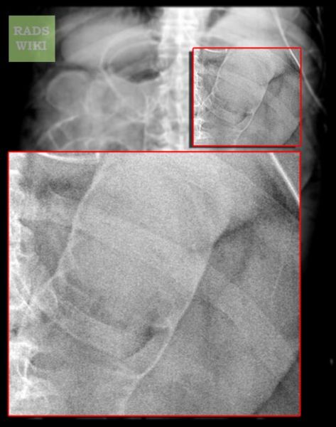
- Rigler’s sign (gas outlining both mucosal and serosal surfaces of bowel wall)
- Falciform ligament sign (gas outlining the falciform ligament)
- Football sign (gas outlining the peritoneal cavity)
-
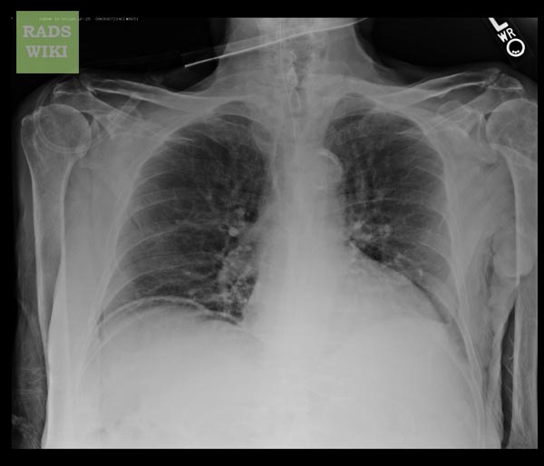
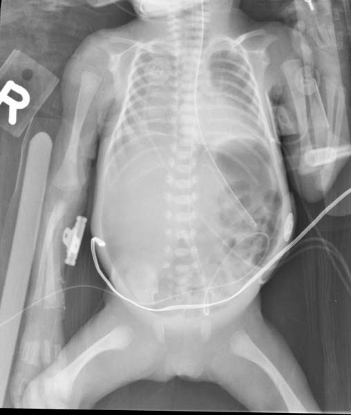
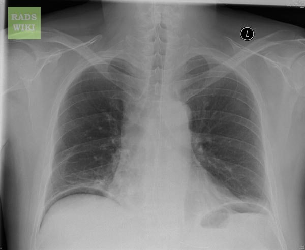 2 days post exploratory laporatomy patient#1 Image courtesy of RadsWiki and copylefted
 -
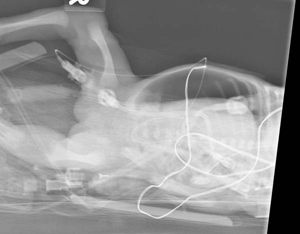
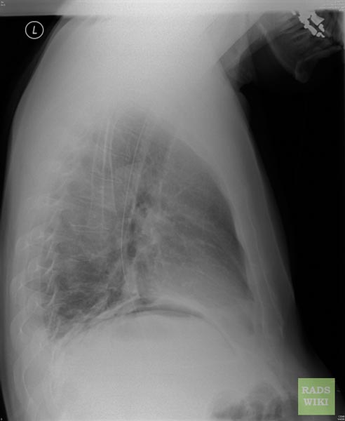 2 days post exploratory laporatomy patient#1 Image courtesy of RadsWiki and copylefted


-
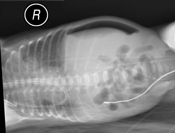
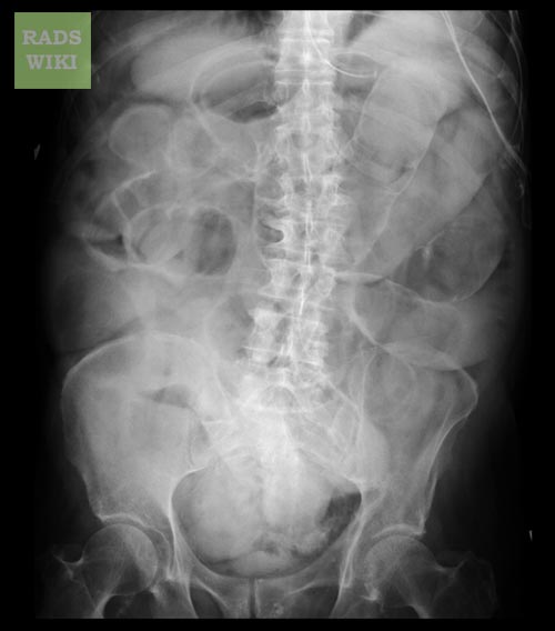 Rigler’s sign patient#2 Image courtesy of RadsWiki and copylefted
 -
 Rigler’s sign patient#2 Image courtesy of RadsWiki and copylefted
 -
 Rigler’s sign patient#2 Image courtesy of RadsWiki and copylefted


-
 patient#3 Image courtesy of RadsWiki and copylefted
 -
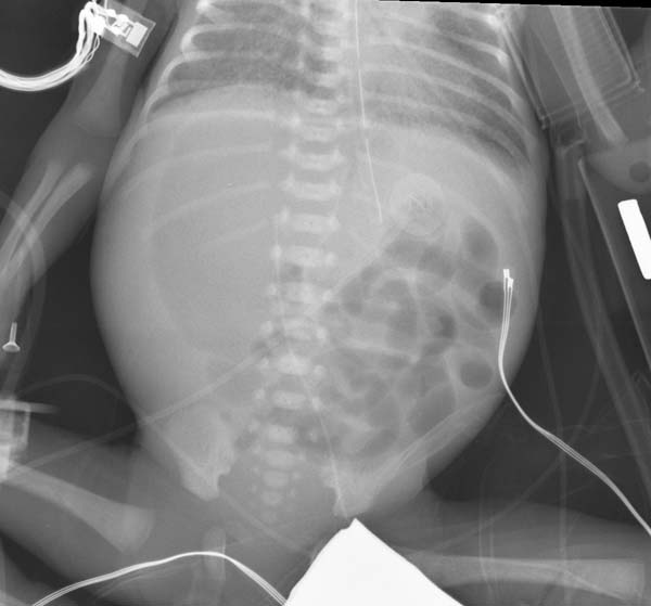 patient#3 Image courtesy of RadsWiki and copylefted
 -
 patient#3 Image courtesy of RadsWiki and copylefted


-
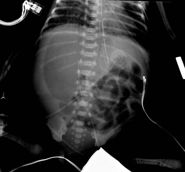
 Large pneumoperitoneum after indomethicin treatment patient#4 Image courtesy of RadsWiki and copylefted
 -
 Large pneumoperitoneum after indomethicin treatment patient#4 Image courtesy of RadsWiki and copylefted


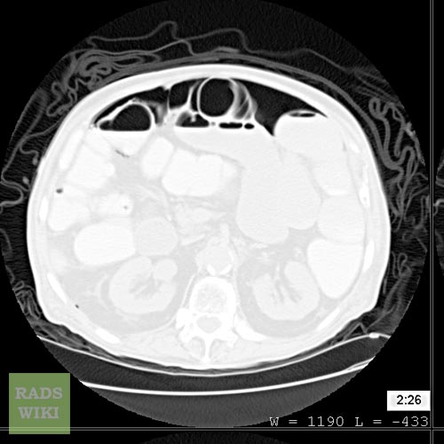
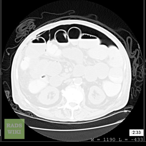
CT
CT can visualize quantities as small as 5 cm³ of air or gas.
-
 Rigler’s sign patient#2 Image courtesy of RadsWiki and copylefted
 -
 Rigler’s sign patient#2 Image courtesy of RadsWiki and copylefted


References
References
Looking for the patient version?
© 2026 MyEClinic – IFTM Institut für Telematik in der Medizin GmbH