
Pulmonary atresia classification with ventricular septal defect
Editor-In-Chief: C. Michael Gibson, M.S., M.D. [1]; Associate Editor(s)-In-Chief: Priyamvada Singh, M.B.B.S. [2], Cafer Zorkun, M.D., Ph.D. [3]; Assistant Editor(s)-In-Chief: Kristin Feeney, B.S. [4]
Overview
Overview
Pulmonary atresia with ventricular septal defect is a congenital lesion associated with the underdevelopment of the right ventricular. Of all congenital cardiac malformations, pulmonary atresia is estimated to occur in 2.5-3.4% of these births.
Pulmonary atresia with ventricular septal defect (PA-VASD)
Pulmonary atresia with ventricular septal defect (PA-VASD)
Pulmonary atresia-sub type VSD is characterized by the opening in the ventricular wall that served as a pathway for the blood flow from the right ventricle to the left ventricle by passing the pulmonary artery due to atresia of the pulmonary artery.
Symptoms severity is widely dependent on the size of the VSD. 50% of the patient also has concomitant ASD due to abnormal migration of the neural crest cell. Pulmonary atresia with VSD is considered one of the most severe variant of the Tetralogy of Fallot, in the which right ventricular outflow in completely blocked due to atresia and represents the 5-10% of the Tetratolgy of Fallot patients. [5][6]
Survival in these patients is dependent on the degree of aorticopulmonary circulation. They are classified as Type A, Type B and Type C on the basic of the where they get there pulmonary blood flow.
Type A would be when the native pulmonary artery supplies the pulmonary circulation via Ductus arteriosus.
Type B would be when native pulmonary arteries receive circulation via major aorticopulmonary vessel or Ductus arteriosus.
Type C would be when the aorticopulmonary collaterals arteries supplies the pulmonary circulation in the absence of the native pulmonary artery. [7]
Echocardiographic finding usually shows mild hypertrophy and dilatation of the RV ( sometimes right atrium ) due to much stain on the RV compared to the atrophy of the RV in PA-IVS.
Images shown below is courtesy of Professor Peter Anderson DVM PhD and published with permission. © PEIR, University of Alabama at Birmingham, Department of Pathology
-
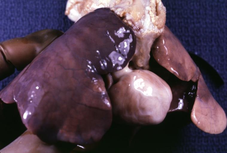 Only an aorta can be seen originating from this pathology specimen. No pulmonary artery is present.

Looking for the patient version?
© 2026 MyEClinic – IFTM Institut für Telematik in der Medizin GmbH