
Pulmonary nodule chest x ray
Editor-In-Chief: C. Michael Gibson, M.S., M.D. [1]Associate Editor(s)-in-Chief: Maria Fernanda Villarreal, M.D. [2]
Overview
Overview
On conventional radiography, characteristic findings of solitary pulmonary nodule, include: well-defined, small, rounded capacities within the pulmonary interstitium, usually 8 mm in diameter, normally surrounded by normal aerated lung.
Chest X Ray
Chest X Ray
- Conventional chest radiograph may be helpful in the diagnosis of pulmonary nodules.
- The majority of pulmonary nodules require further evaluation with CT scan and MRI
- On conventional radiography, characteristic findings of pulmonary nodule, include:[1]
- Soft-tissue density mass
- Round or oval in shape
- Smooth margin
- Diameter of 8 mm and irregular margins
- Surrounded by areas of ground glass change
Gallery
Gallery
-
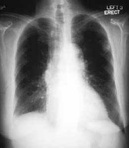 Malignant pulmonary nodule:The patient is a 67 year old woman with a solitary pulmonary nodule on a recent chest x-ray. A retrospective review of prior chest x-rays suggests that this is nodule is of recent origin. This lesion was felt to be too peripheral for reliable bronchial wash findings Images shown above are courtesy of Professor Peter Anderson DVM PhD and published with permission © PEIR, University of Alabama at Birmingham, Department of Pathology
 -
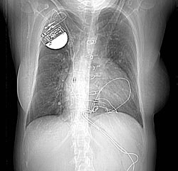 Arteriovenous malformations: Two pulmonary arteriovenous malformations consistent with the nodules seen on the recent chest film. There is breathing artifact on several of the images and other tiny AVMs cannot be excluded.Images shown above are courtesy of Professor Peter Anderson DVM PhD and published with permission © PEIR, University of Alabama at Birmingham, Department of Pathology


References
References
- ↑ Kundel HL (1981). “Predictive value and threshold detectability of lung tumors”. Radiology. 139 (1): 25–9. doi:10.1148/radiology.139.1.7208937. PMID 7208937.
Looking for the patient version?
© 2026 MyEClinic – IFTM Institut für Telematik in der Medizin GmbH