

Health Dictionary
Reliable, easy-to-read patient information on common medical conditions — written in plain language, free of jargon, and grounded in current evidence. Browse the most frequent diagnoses, understand what they mean for everyday life, and learn which steps and questions help you have a focused conversation with your doctor.
Renal artery stenosis MRI
Introduction
Overview
Overview
Diagnosis by magnetic resonance angiography (MRA) is considered class I recommendation. Gadolinium-based MRA has less nephrotoxic characterstics with good visualization of the renal arteries and perirenal pathologies. Presence of previous stents or metallic objects are considered a contraindication for the use of MRA.[1]
MRI
MRI
There are no MRI findings associated with [disease name].
OR
[Location] MRI may be helpful in the diagnosis of [disease name]. Findings on MRI suggestive of/diagnostic of [disease name] include:
- [Finding 1]
- [Finding 2]
- [Finding 3]
OR
There are no MRI findings associated with [disease name]. However, a MRI may be helpful in the diagnosis of complications of [disease name], which include:
- [Complication 1]
- [Complication 2]
- [Complication 3]
Case #1
-
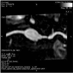 High grade left main renal artery stenosis, MRA. 76 year old male with hypertension.
 -
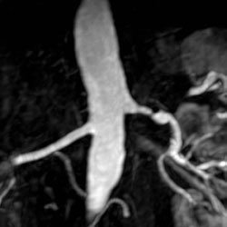 High grade left main renal artery stenosis, MRA. 76 year old male with hypertension.
 -
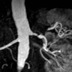 High grade left main renal artery stenosis, MRA. 76 year old male with hypertension.
 -
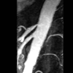 High grade left main renal artery stenosis, MRA. 76 year old male with hypertension.
 -
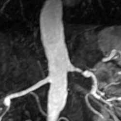 High grade left main renal artery stenosis, MRA. 76 year old male with hypertension.
 -
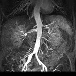 High grade left main renal artery stenosis, MRA. 76 year old male with hypertension.
 -
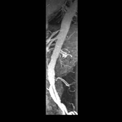 High grade left main renal artery stenosis, MRA. 76 year old male with hypertension.
 -
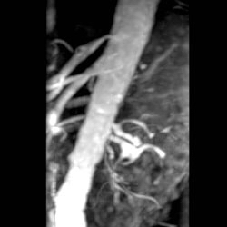 High grade left main renal artery stenosis, MRA. 76 year old male with hypertension.
 -
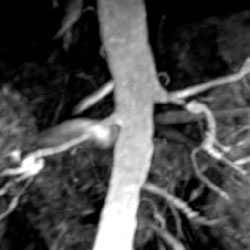 High grade left main renal artery stenosis, MRA. 76 year old male with hypertension.


Case #2
-
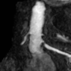 MRA: Renal artery stenosis.
 -
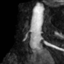 MRA: Renal artery stenosis.
 -
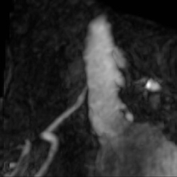 MRA: Renal artery stenosis.
 -
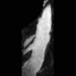 MRA: Renal artery stenosis.
 -
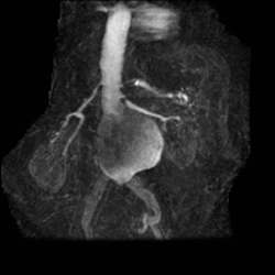 MRA: Renal artery stenosis.
 -
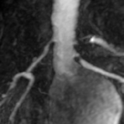 MRA: Renal artery stenosis.
 -
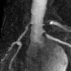 MRA: Renal artery stenosis.
 -
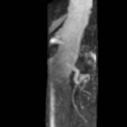 MRA: Renal artery stenosis.
 -
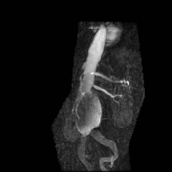 MRA: Renal artery stenosis.
 -
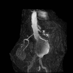 MRA: Renal artery stenosis.


Case #3
-
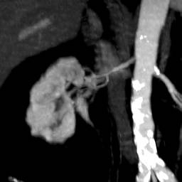 Atherosclerotic plaques seen in the distal aortic arch, aortic bifurcation, and in the external iliac arteries. right renal artery stenosis. A 74 year-old male who is status post thromboembolic event of his RLE.
 -
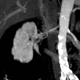 Atherosclerotic plaques seen in the distal aortic arch, aortic bifurcation, and in the external iliac arteries. right renal artery stenosis. A 74 year-old male who is status post thromboembolic event of his RLE.
 -
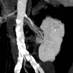 Atherosclerotic plaques seen in the distal aortic arch, aortic bifurcation, and in the external iliac arteries. right renal artery stenosis. A 74 year-old male who is status post thromboembolic event of his RLE.
 -
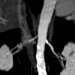 Atherosclerotic plaques seen in the distal aortic arch, aortic bifurcation, and in the external iliac arteries. right renal artery stenosis. A 74 year-old male who is status post thromboembolic event of his RLE.
 -
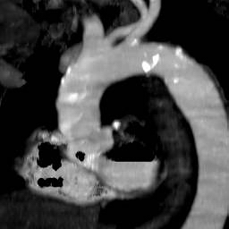 Atherosclerotic plaques seen in the distal aortic arch, aortic bifurcation, and in the external iliac arteries. right renal artery stenosis. A 74 year-old male who is status post thromboembolic event of his RLE.
 -
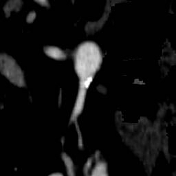 Atherosclerotic plaques seen in the distal aortic arch, aortic bifurcation, and in the external iliac arteries. right renal artery stenosis. A 74 year-old male who is status post thromboembolic event of his RLE.
 -
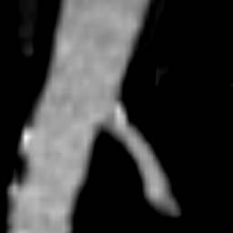 Atherosclerotic plaques seen in the distal aortic arch, aortic bifurcation, and in the external iliac arteries. right renal artery stenosis. A 74 year-old male who is status post thromboembolic event of his RLE.
 -
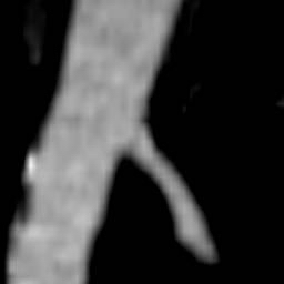 Atherosclerotic plaques seen in the distal aortic arch, aortic bifurcation, and in the external iliac arteries. right renal artery stenosis. A 74 year-old male who is status post thromboembolic event of his RLE.
 -
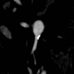 Atherosclerotic plaques seen in the distal aortic arch, aortic bifurcation, and in the external iliac arteries. right renal artery stenosis. A 74 year-old male who is status post thromboembolic event of his RLE.
 -
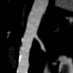 Atherosclerotic plaques seen in the distal aortic arch, aortic bifurcation, and in the external iliac arteries. right renal artery stenosis. A 74 year-old male who is status post thromboembolic event of his RLE.
 -
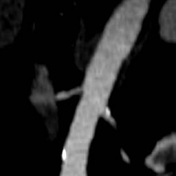 Atherosclerotic plaques seen in the distal aortic arch, aortic bifurcation, and in the external iliac arteries. right renal artery stenosis. A 74 year-old male who is status post thromboembolic event of his RLE.
 -
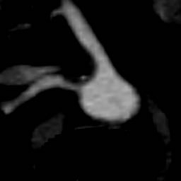 Atherosclerotic plaques seen in the distal aortic arch, aortic bifurcation, and in the external iliac arteries. right renal artery stenosis. A 74 year-old male who is status post thromboembolic event of his RLE.
 -
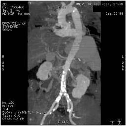 Atherosclerotic plaques seen in the distal aortic arch, aortic bifurcation, and in the external iliac arteries. right renal artery stenosis. A 74 year-old male who is status post thromboembolic event of his RLE.
 -
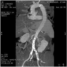 Atherosclerotic plaques seen in the distal aortic arch, aortic bifurcation, and in the external iliac arteries. right renal artery stenosis. A 74 year-old male who is status post thromboembolic event of his RLE.
 -
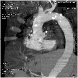 Atherosclerotic plaques seen in the distal aortic arch, aortic bifurcation, and in the external iliac arteries. right renal artery stenosis. A 74 year-old male who is status post thromboembolic event of his RLE.
 -
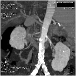 Atherosclerotic plaques seen in the distal aortic arch, aortic bifurcation, and in the external iliac arteries. right renal artery stenosis. A 74 year-old male who is status post thromboembolic event of his RLE.
 -
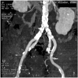 Atherosclerotic plaques seen in the distal aortic arch, aortic bifurcation, and in the external iliac arteries. right renal artery stenosis. A 74 year-old male who is status post thromboembolic event of his RLE.
 -
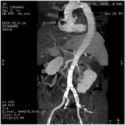 Atherosclerotic plaques seen in the distal aortic arch, aortic bifurcation, and in the external iliac arteries. right renal artery stenosis. A 74 year-old male who is status post thromboembolic event of his RLE.


Cardiac MRI in Renal artery stenosis
ACC/AHA Guidelines- ACCF/ACR/AHA/NASCI/SCMR 2010 Expert Consensus Document on Cardiovascular Magnetic Resonance[2] (DO NOT EDIT)
| “ |
CMR may be used for evaluating renal arterial stenoses and quantifying renal arterial blood flow. CE-MRA indicates contrast-enhanced magnetic resonance angiography; CMR, cardiovascular magnetic resonance; ECG, electrocardiogram; LGE, late gadolinium enhancement; LV, left ventricular; RV, right ventricular; MRA, magnetic resonance angiography; and PAD, peripheral arterial |
” |
References
References
- ↑ Hirsch AT, Haskal ZJ, Hertzer NR, Bakal CW, Creager MA, Halperin JL; et al. (2006). “ACC/AHA 2005 guidelines for the management of patients with peripheral arterial disease (lower extremity, renal, mesenteric, and abdominal aortic): executive summary a collaborative report from the American Association for Vascular Surgery/Society for Vascular Surgery, Society for Cardiovascular Angiography and Interventions, Society for Vascular Medicine and Biology, Society of Interventional Radiology, and the ACC/AHA Task Force on Practice Guidelines (Writing Committee to Develop Guidelines for the Management of Patients With Peripheral Arterial Disease) endorsed by the American Association of Cardiovascular and Pulmonary Rehabilitation; National Heart, Lung, and Blood Institute; Society for Vascular Nursing; TransAtlantic Inter-Society Consensus; and Vascular Disease Foundation”. J Am Coll Cardiol. 47 (6): 1239–312. doi:10.1016/j.jacc.2005.10.009. PMID 16545667.
- ↑ American College of Cardiology Foundation Task Force on Expert Consensus Documents. Hundley WG, Bluemke DA, Finn JP, Flamm SD, Fogel MA; et al. (2010). “ACCF/ACR/AHA/NASCI/SCMR 2010 expert consensus document on cardiovascular magnetic resonance: a report of the American College of Cardiology Foundation Task Force on Expert Consensus Documents”. Circulation. 121 (22): 2462–508. doi:10.1161/CIR.0b013e3181d44a8f. PMC 3034132. PMID 20479157.
Editor-In-Chief: C. Michael Gibson, M.S., M.D. [1]