
Renal artery stenosis ultrasound
Editor-In-Chief: C. Michael Gibson, M.S., M.D. [1]
Overview
Overview
Duplex ultrasonography is a first line non-invasive imaging technique for the diagnosis of atherosclerotic renal artery stenosis.
Duplex Ultrasonography
Duplex Ultrasonography
Diagnosis by Duplex ultrasonography is considered class I recommendation. It may be used as an initial screening tool for diagnosis of atherosclerotic renal artery stenosis. Ultrasonography might not be very accurate in obese patients or those intestinal gas.[1]
-
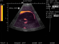
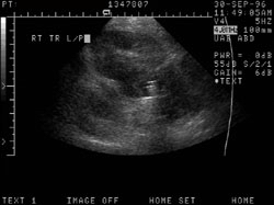 Abdominal pain in a patient with previous renal transplant. 1) Juxtanephric mass surrounding renal vessels, differential includes adenopathy, hematoma and abscess. 2) Renal artery stenosis of CRT.
 -
 Abdominal pain in a patient with previous renal transplant. 1) Juxtanephric mass surrounding renal vessels, differential includes adenopathy, hematoma and abscess. 2) Renal artery stenosis of CRT


Doppler Ultrasonography
Case #1
-
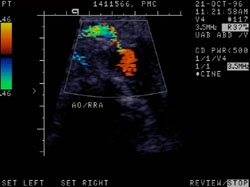
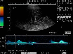
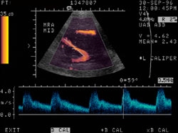
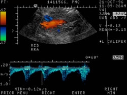 Renal artery stenosis. The patient is a 75 year old white female with a history of hypertension. she is s/p left nephrectomy approximately 10 years ago for renal cell carcinoma. 1. Definite renal artery stenosis involving the origin of the right renal artery. 2. Focal dilatation of the distal abdominal aorta, but no frank aneurysm.
 -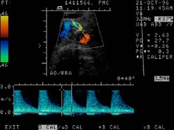
 Renal artery stenosis. The patient is a 75 year old white female with a history of hypertension. she is s/p left nephrectomy approximately 10 years ago for renal cell carcinoma. 1. Definite renal artery stenosis involving the origin of the right renal artery. 2. Focal dilatation of the distal abdominal aorta, but no frank aneurysm.


Case #2
-
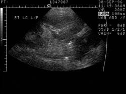
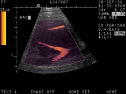 Abdominal pain in a patient with previous renal transplant. 1) Juxtanephric mass surrounding renal vessels, differential includes adenopathy, hematoma and abscess. 2) Renal artery stenosis of CRT
 -
 Abdominal pain in a patient with previous renal transplant. 1) Juxtanephric mass surrounding renal vessels, differential includes adenopathy, hematoma and abscess. 2) Renal artery stenosis of CRT
 -
 Abdominal pain in a patient with previous renal transplant. 1) Juxtanephric mass surrounding renal vessels, differential includes adenopathy, hematoma and abscess. 2) Renal artery stenosis of CRT
 -
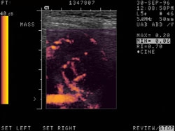 Abdominal pain in a patient with previous renal transplant. 1) Juxtanephric mass surrounding renal vessels, differential includes adenopathy, hematoma and abscess. 2) Renal artery stenosis of CRT


References
References
- ↑ Anderson JL, Halperin JL, Albert NM, Bozkurt B, Brindis RG, Curtis LH; et al. (2013). “Management of patients with peripheral artery disease (compilation of 2005 and 2011 ACCF/AHA guideline recommendations): a report of the American College of Cardiology Foundation/American Heart Association Task Force on Practice Guidelines”. Circulation. 127 (13): 1425–43. doi:10.1161/CIR.0b013e31828b82aa. PMID 23457117.
Looking for the patient version?
© 2026 MyEClinic – IFTM Institut für Telematik in der Medizin GmbH