
Renal ectopia ultrasound
Editor-In-Chief: C. Michael Gibson, M.S., M.D. [1]; Associate Editor(s)-in-Chief:
Overview
Overview
There are no echocardiography/ultrasound findings associated with [disease name].
OR
Echocardiography/ultrasound may be helpful in the diagnosis of [disease name]. Findings on an echocardiography/ultrasound suggestive of/diagnostic of [disease name] include [finding 1], [finding 2], and [finding 3].
OR
There are no echocardiography/ultrasound findings associated with [disease name]. However, an echocardiography/ultrasound may be helpful in the diagnosis of complications of [disease name], which include [complication 1], [complication 2], and [complication 3].
Echocardiography/Ultrasound
Echocardiography/Ultrasound
There are no echocardiography/ultrasound findings associated with [disease name].
OR
Echocardiography/ultrasound may be helpful in the diagnosis of [disease name]. Findings on an echocardiography/ultrasound suggestive of/diagnostic of [disease name] include:
- [Finding 1]
- [Finding 2]
- [Finding 3]
OR
There are no echocardiography/ultrasound findings associated with [disease name]. However, an echocardiography/ultrasound may be helpful in the diagnosis of complications of [disease name], which include:
- [Complication 1]
- [Complication 2]
- [Complication 3]
CT Scan
CT Scan
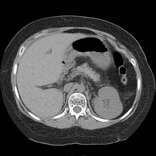
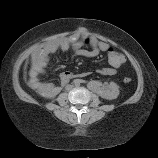
Findings on CT should be interpreted in light of bowel laxity in the region of the empty renal fossa. In particular, distinction must be made from internal hernia. Patient #1: CT images demonstrate a pelvic kidney


Patient #1: CT images demonstrate cross-fused renal ectopia
-
 Kidney 1
 -

-
 Kidney 2


Patient #2: Renal scan images demonstrate cross-fused renal ectopia


Looking for the patient version?
© 2026 MyEClinic – IFTM Institut für Telematik in der Medizin GmbH