
Salter-Harris fractures
Editor-In-Chief: C. Michael Gibson, M.S., M.D. [1]; Associate Editor(s)-in-Chief: Prashanth Saddala M.B.B.S
Overview
Overview
Salter-Harris fractures are descriptive terms for fractures affecting the growth plate of a bone. Once bone growth has completed, the term “Salter-Harris Fracture” no longer applies.
Classification
Classification
There are six types of Salter-Harris fractures:[1]
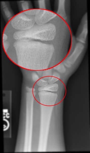
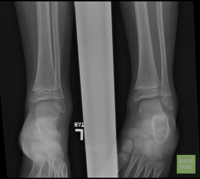
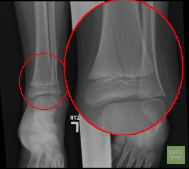
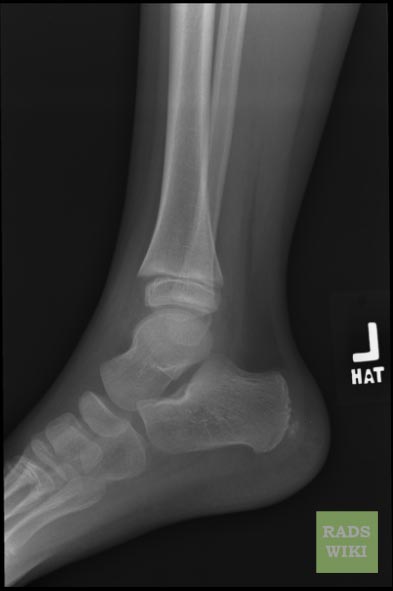
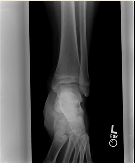
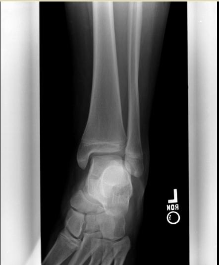
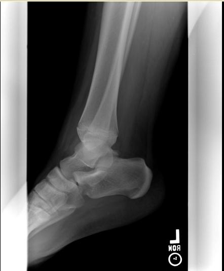
- Type I – A transverse fracture through the growth plate (also referred to as the “physis”):[2] 6% incidence
-
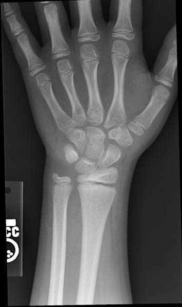 Salter-Harris fracture-I Image courtesy of RadsWiki and copylefted
 -
 Salter-Harris fracture-I Image courtesy of RadsWiki and copylefted


- Type II – A fracture through the growth plate and the metaphysis, sparing the epiphysis:[3] 75% incidence
-
 Salter-Harris fracture-II Image courtesy of RadsWiki and copylefted
 -
 Salter-Harris fracture-II Image courtesy of RadsWiki and copylefted
 -
 Salter-Harris fracture-II Image courtesy of RadsWiki and copylefted


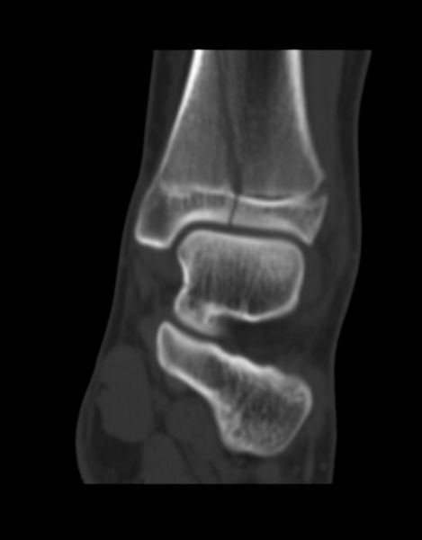
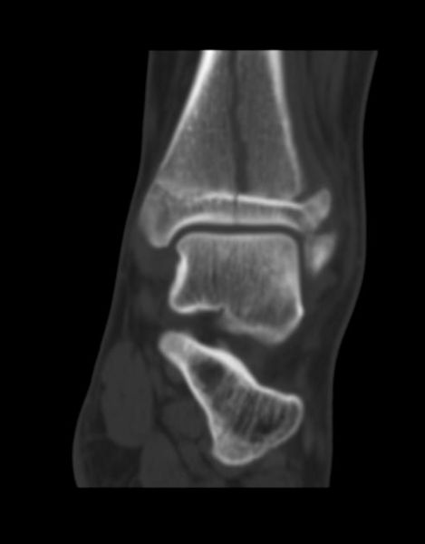
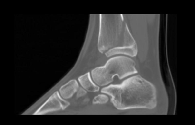
- Type III – A fracture through growth plate and epiphysis, sparing the metaphysis:[4] 8% incidence
-
 Salter-Harris fracture-III Image courtesy of RadsWiki and copylefted
 -
 Salter-Harris fracture-III Image courtesy of RadsWiki and copylefted
 -
 Salter-Harris fracture-III Image courtesy of RadsWiki and copylefted
 -
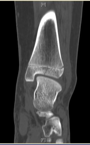 Salter-Harris fracture-III Image courtesy of RadsWiki and copylefted
 -
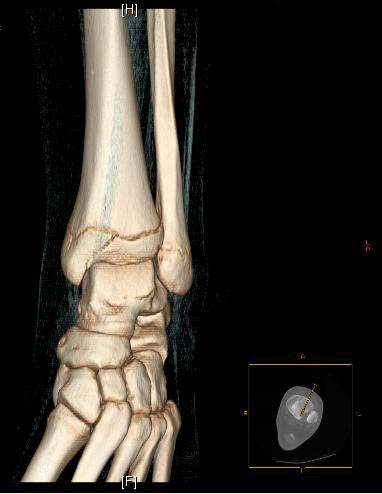 Salter-Harris fracture-III Image courtesy of RadsWiki and copylefted
 -
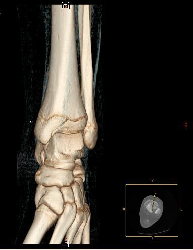 Salter-Harris fracture-III Image courtesy of RadsWiki and copylefted
 -
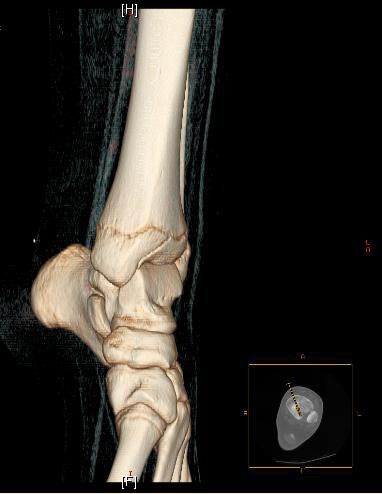 Salter-Harris fracture-III Image courtesy of RadsWiki and copylefted


- Type IV – A fracture through all three elements of the bone, the growth plate, metaphysis, and epiphysis:[5] 10% incidence
-
 Salter-Harris fracture-IV Image courtesy of RadsWiki and copylefted
 -
 Salter-Harris fracture-IV Image courtesy of RadsWiki and copylefted
 -
 Salter-Harris fracture-IV Image courtesy of RadsWiki and copylefted


- Type V – A compression fracture of the growth plate (resulting in a decrease in the perceived space between the epiphysis and diaphysis on x-ray):[6] 1% incidence
- Type VI – Injury to the peripheral portion of the physis and a resultant bony bridge formation which my produce an angular deformity. (Added in 1969 by Mercer Rang.)
References
References
- ↑ Template:Chorus
- ↑ “S.H. Type I – Wheeless’ Textbook of Orthopaedics”.
- ↑ “S.H. Type II – Wheeless’ Textbook of Orthopaedics”.
- ↑ “Salter Harris Type III Frx – Wheeless’ Textbook of Orthopaedics”.
- ↑ “Salter Harris: Type IV – Wheeless’ Textbook of Orthopaedics”.
- ↑ “Type V – Wheeless’ Textbook of Orthopaedics”.
Looking for the patient version?
© 2026 MyEClinic – IFTM Institut für Telematik in der Medizin GmbH