

Health Dictionary
Reliable, easy-to-read patient information on common medical conditions — written in plain language, free of jargon, and grounded in current evidence. Browse the most frequent diagnoses, understand what they mean for everyday life, and learn which steps and questions help you have a focused conversation with your doctor.
Sclera
Introduction
The sclera is the opaque (usually white), fibrous, protective layer of the eye containing collagen and elastic fibers.[1] In children, it is thinner and shows some of the underlying pigment, appearing slightly blue. In the old, however, fatty deposits on the sclera can make it appear slightly yellow.
The sclera forms the posterior five sixths of the connective tissue coat of the globe. The sclera maintains the shape of the globe, offering resistance to internal and external forces, and provides an attachment for the extraocular muscle insertions. The thickness of the sclera varies from 1mm at the posterior pole to 0.3 mm just behind the rectus muscle insertions.
Additional images
Additional images
-
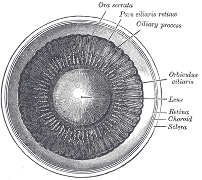 Interior of anterior half of bulb of eye.
 -
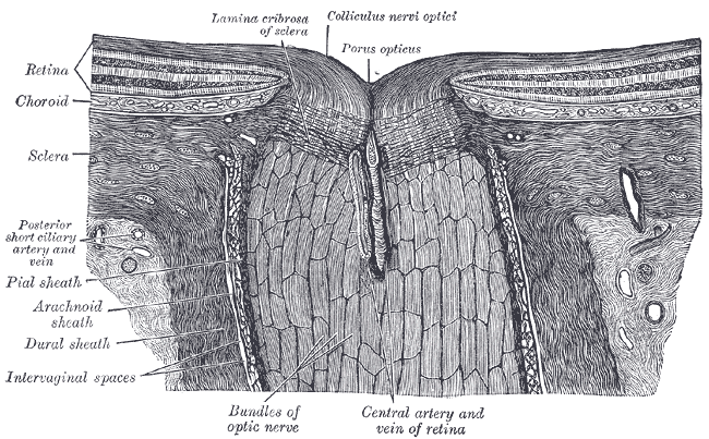 The terminal portion of the optic nerve and its entrance into the eyeball, in horizontal section.
 -
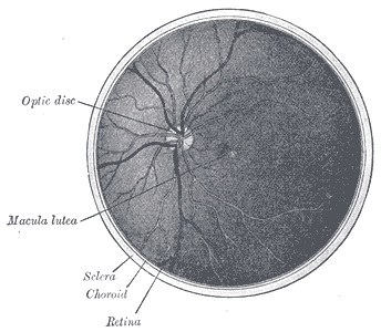 The interior of the posterior half of the left eyeball.


References
References
- ↑ Cassin, B. and Solomon, S. Dictionary of Eye Terminology. Gainesville, Florida: Triad Publishing Company, 1990.
External links
External links
- Histology image: 08008loa – Histology Learning System at Boston University
- Template:UMichAtlas – “Sagittal Section Through the Eyeball”
Template:Eye
Template:Visual system
Template:Muscles of orbit
ca:Escleròtica de:Sclera it:Sclera lt:Odena nl:Sclera no:Sklera sk:Očné bielko sv:senhinnan
Editor-In-Chief: Template:Infobox Anatomy C. Michael Gibson, M.S., M.D. [1]