
Submucosa
Editor-In-Chief: C. Michael Gibson, M.S., M.D. [1]
Overview
Overview
In the gastrointestinal tract, the submucosa is the layer of loose connective tissue that supports the mucosa, as well as joins the mucosa to the bulk of underlying smooth muscle (fibers running circularly within layer of longitudinal muscle).
Contents
Contents
Blood vessels, lymphatic vessels, and nerves (all supplying the mucosa) will run through here.
Tiny parasympathetic ganglia are scattered around forming the submucosal plexus (or “Meissner’s plexus”) where preganglionic parasympathetic neurons synapse with postganglionic nerve fibers that supply the muscularis mucosae.
The submucosa in endoscopy
The submucosa in endoscopy
Identification of the submucosa plays an important role in diagnostic and therapeutic endoscopy, where special fibre-optic cameras are used to perform procedures on the gastrointestinal tract. Abnormalities of the submucosa, such as gastrointestinal stromal tumors, usually show integrity of the mucosal surface.
The submucosa is also identified in endoscopic ultrasound to identify the depth of tumours and to identify other abnormalities. An injection of dye, saline, or epinephrine into the submucosa is imperative in the safe removal of certain polyps.
Endoscopic mucosal resection involves removal of the mucosal layer, and in order to be done safely, a submucosal injection of dye is performed to ensure integrity at the beginning of the procedure.
Additional images
Additional images
-
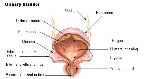 Bladder.
 -
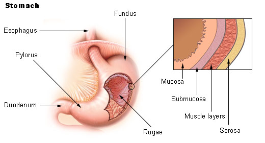 Stomach.
 -
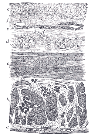 Section of the human esophagus. Moderately magnified.
 -
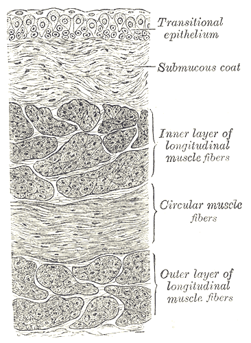 Vertical section of bladder wall.
 -
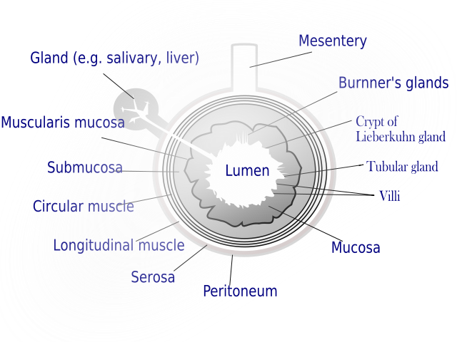 General structure of the gut wall showing the submucosa.


External links
External links
- Template:UCDavisOrganology – “Mammal, whole system (LM, Low)”
- Template:EMedicineDictionary
- Histology image: 00102loa – Histology Learning System at Boston University – “Tissues, Layers, and Organs: transverse section of rat gut”
- Essentials of Human Physiology by Thomas M. Nosek. Section 6/6ch1/s6ch1_9.
- Template:OklahomaHistology
- Template:AnatomyAtlasesMicroscopic – “Duodenum and Jejunum”
- Histology at edteched.uottawa.ca
Looking for the patient version?
© 2026 MyEClinic – IFTM Institut für Telematik in der Medizin GmbH