

Health Dictionary
Reliable, easy-to-read patient information on common medical conditions — written in plain language, free of jargon, and grounded in current evidence. Browse the most frequent diagnoses, understand what they mean for everyday life, and learn which steps and questions help you have a focused conversation with your doctor.
Superior vena cava syndrome CT
Introduction
Overview
Overview
On enhanced CT scan, findings include location and severity of the superior vena cava obstruction, superimposed thrombosis, a mediastinal mass or lymphadenopathy, collateral vessels, and associated lung masses. CT scan is the imaging modality of choice.
CT
CT
- CT scan is the imaging modality of choice for the diagnosis of superior vena cava syndrome.
- On CT scan, superior vena cava syndrome is characterized by:[1]
- Location and severity of the superior vena cava obstruction
- Superimposed thrombosis
- Mediastinal mass or lymphadenopathy
- Collateral vessels and associated lung masses
Gallery
Gallery
-
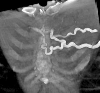 Collaterals on CT scan in a patient with superior vena cava syndrome

-
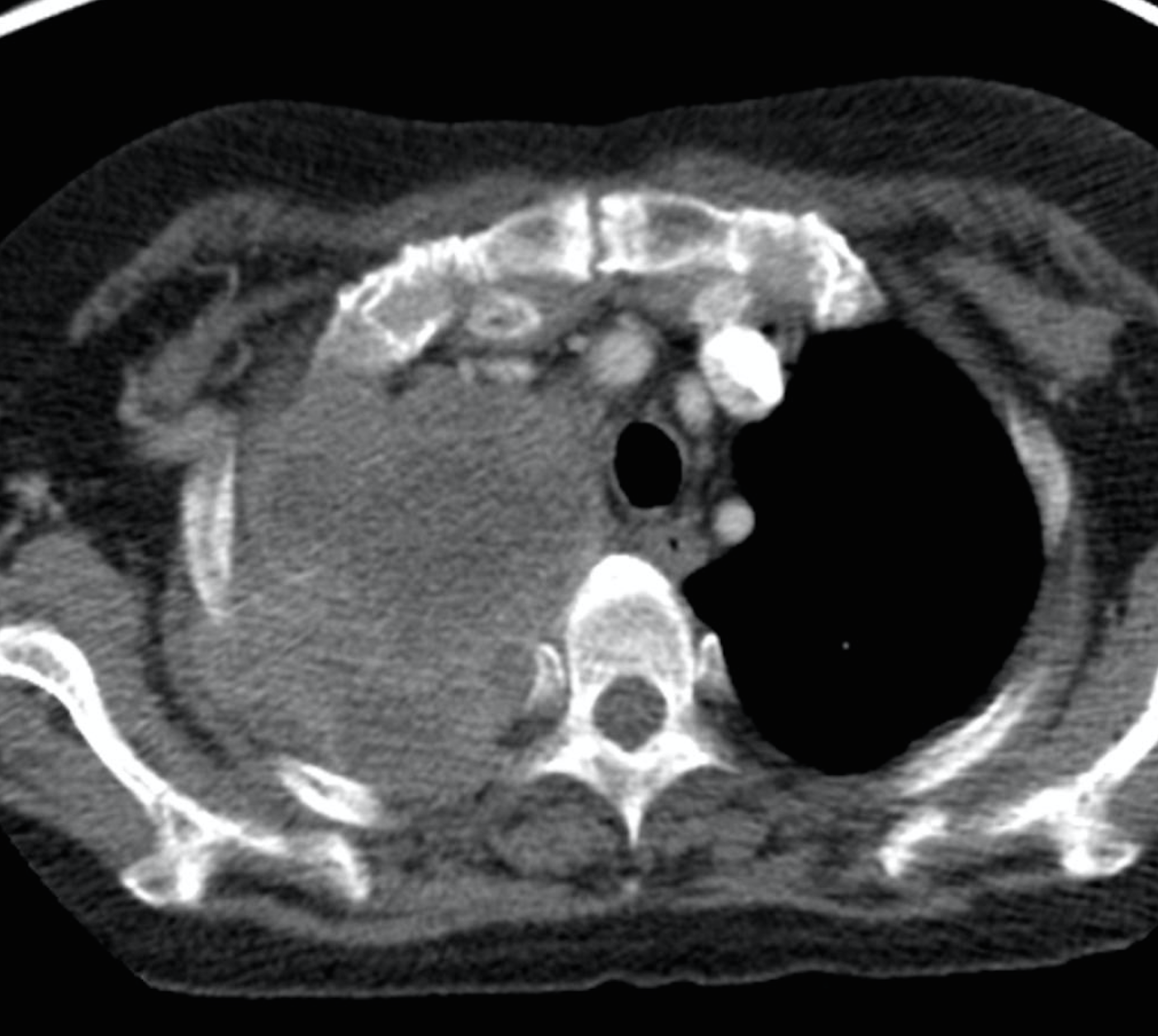 Lung cancer with vena cava invasion.

References
References
- ↑ Superior Vena Cava Syndrome.Dr Amir Rezaee and Radswiki et al. Radiopedia http://radiopaedia.org/articles/superior-vena-cava-obstruction Accessed on January 13, 2016
Editor-In-Chief: C. Michael Gibson, M.S., M.D. [1]Associate Editor(s)-in-Chief: Maria Fernanda Villarreal, M.D. [2]