

Health Dictionary
Reliable, easy-to-read patient information on common medical conditions — written in plain language, free of jargon, and grounded in current evidence. Browse the most frequent diagnoses, understand what they mean for everyday life, and learn which steps and questions help you have a focused conversation with your doctor.
Temporalis muscle
Introduction
Structure
Structure
It arises from the temporal fossa and the deep part of temporal fascia. It passes medial to the zygomatic arch and inserts onto the coronoid process of the mandible.
The temporalis muscle is covered by the temporal fascia, also known as the temporal aponeurosis.
The muscle can be felt if one places their fingers on their temples (on the sides of their head, just behind the eyebrows), while clenching and unclenching their teeth.
Innervation
Innervation
As with the other muscles of mastication, control of the temporalis muscle comes from the third (mandibular) branch of the trigeminal nerve. Specifically, the temporalis is innervated by the deep temporal nerves.
Actions
Actions
Contraction of the temporalis muscle elevates the mandible. The somewhat horizontal fibers of the posterior part of the muscle retract the mandible.
Additional images
Additional images
-
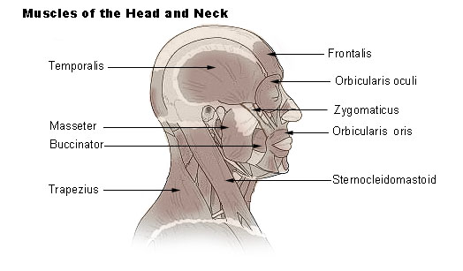 Muscles of head and neck
 -
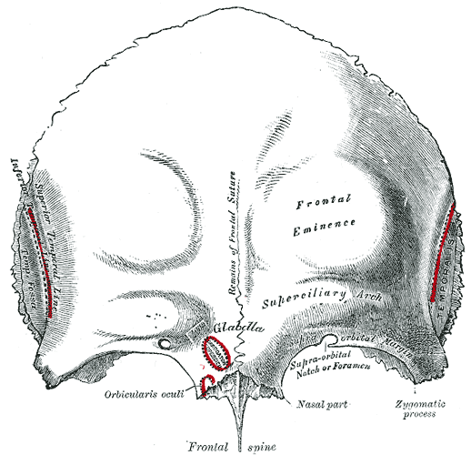 Frontal bone. Outer surface.
 -
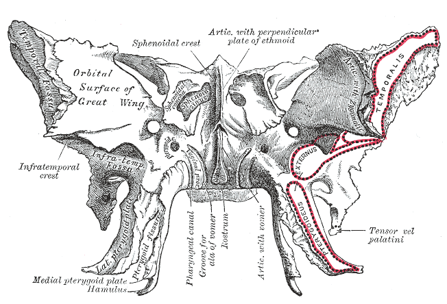 Sphenoid bone. Anterior and inferior surfaces.
 -
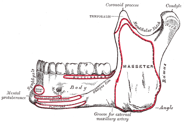 Mandible. Outer surface. Side view.
 -
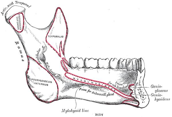 Mandible. Inner surface. Side view.
 -
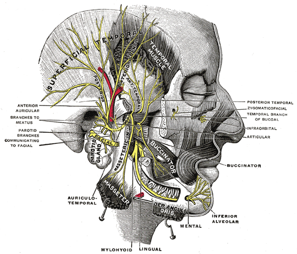 Mandibular division of the trifacial nerve.


External links
External links
- Template:MuscleLoyola
- Template:SUNYAnatomyLabs – “Infratemporal Fossa: The Temporalis Muscle”
Editor-In-Chief: C. Michael Gibson, M.S., M.D. [1]