

Health Dictionary
Reliable, easy-to-read patient information on common medical conditions — written in plain language, free of jargon, and grounded in current evidence. Browse the most frequent diagnoses, understand what they mean for everyday life, and learn which steps and questions help you have a focused conversation with your doctor.
Thromboembolism CT
Introduction
CT
CT
Computed tomography with radiocontrast, effectively a pulmonary angiogram imaged by CT and also known as CT pulmonary angiography (CTPA), is increasingly used as the mainstay in diagnosis. Advantages are clinical equivalence, its non-invasive nature, its greater availability to patients, and the possibility of picking up other lung disorders from the differential diagnosis in case there is no pulmonary embolism.
CT Findings in Acute PE
- Thrombus is located centrally within the vascular lumen or occludes the vessel (vessel cut-off sign)
- Commonly causes distention of the involved vessel.
CT Findings in Chronic PE
- Eccentric and contiguous changes of the vessel wall
- Reduces the arterial diameter by more than 50%
- Evidence of recanalization within the thrombus
- An arterial web is present
-
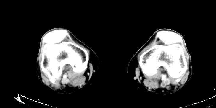 Left leg. Deep venous thrombosis
 -
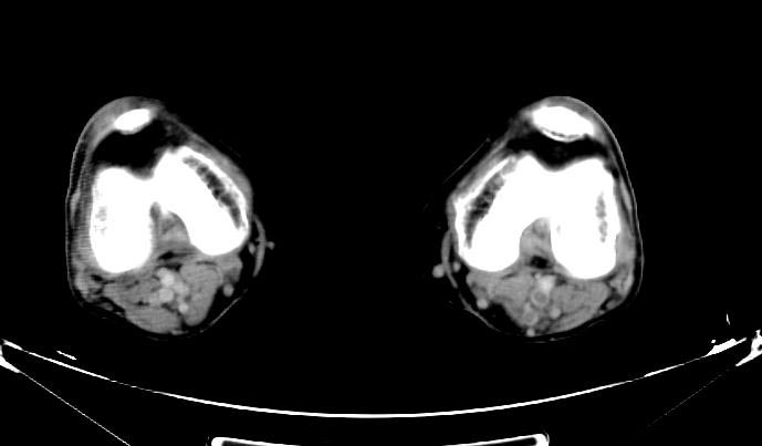 Left leg. Deep venous thrombosis
 -
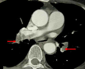 CT pulmonary angiogram. Clots in both the left and the right pulmonary arteries (red arrows). Source


Patient with Shortness of Breath
-
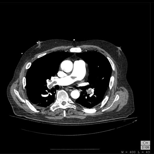 Pulmonary embolism: Patient presented with Shortness of breath
 -
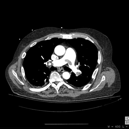 Pulmonary embolism: Patient presented with Shortness of breath
 -
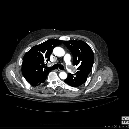 Pulmonary embolism: Patient presented with Shortness of breath
 -
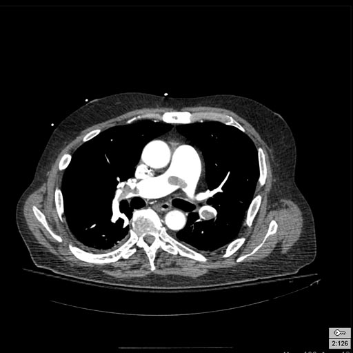 Pulmonary embolism: Patient presented with Shortness of breath


Patient with Acute RBBB
-
 Pulmonary embolism: Patient presented with Acute RBBB
 -
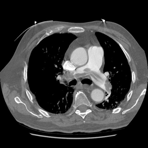 Pulmonary embolism: Patient presented with Acute RBBB


Editor-In-Chief: C. Michael Gibson, M.S., M.D. [1]