
Varicocele echocardiography or ultrasound
Editor-In-Chief: C. Michael Gibson, M.S., M.D. [1]
Overview
Overview
Ultrasound
Ultrasound
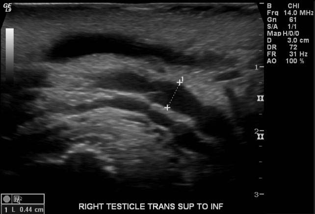
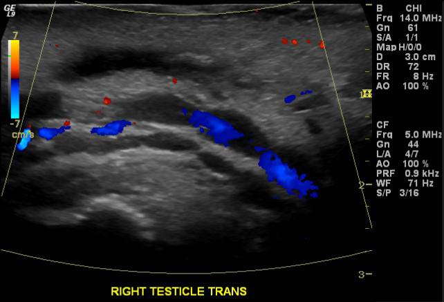
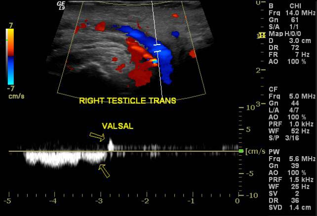
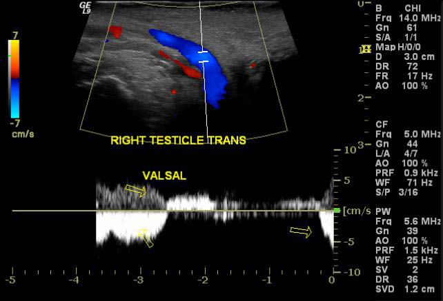
Varicocele can be reliably diagnosed with ultrasound, which will show dilatation of the vessels of the pampiniform plexus to greater than 2 mm. The patient being studied should undergo a provocative maneuver, such as a Valsalva maneuver (straining, like he is trying to have a bowel movement) or standing up during the exam, both of which are designed to increase intra-abdominal venous pressure and increase the dilatation of the veins. Doppler ultrasound is a technique of measuring the speed at which blood is flowing in a vessel. An ultrasound machine that has a Doppler mode can see blood reverse direction in a varicocele with a Valsalva, increasing the sensitivity of the examination.
-
 Varicocele
 -
 Varicocele
 -
 Varicocele
 -
 Varicocele


Looking for the patient version?
© 2026 MyEClinic – IFTM Institut für Telematik in der Medizin GmbH