
Vertebral vein
Editor-In-Chief: C. Michael Gibson, M.S., M.D. [1]
The vertebral vein is formed in the suboccipital triangle, from numerous small tributaries which spring from the internal vertebral venous plexuses and issue from the vertebral canal above the posterior arch of the atlas.
They unite with small veins from the deep muscles at the upper part of the back of the neck, and form a vessel which enters the foramen in the transverse process of the atlas, and descends, forming a dense plexus around the vertebral artery, in the canal formed by the foramina transversaria of the cervical vertebrae.
This plexus ends in a single trunk, which emerges from the foramen transversarium of the sixth cervical vertebra, and opens at the root of the neck into the back part of the innominate vein near its origin, its mouth being guarded by a pair of valves.
On the right side, it crosses the first part of the subclavian artery.
Additional images
Additional images
-
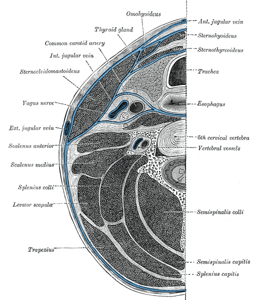 Section of the neck at about the level of the sixth cervical vertebra.

Looking for the patient version?
© 2026 MyEClinic – IFTM Institut für Telematik in der Medizin GmbH