

Health Dictionary
Reliable, easy-to-read patient information on common medical conditions — written in plain language, free of jargon, and grounded in current evidence. Browse the most frequent diagnoses, understand what they mean for everyday life, and learn which steps and questions help you have a focused conversation with your doctor.
Vitelline duct
Introduction
Overview
Overview
At the end of the fourth week the yolk-sac presents the appearance of a small pear-shaped vesicle (umbilical vesicle) opening into the digestive tube by a long narrow tube, the vitelline duct.
The vesicle can be seen in the after-birth as a small, somewhat oval-shaped body whose diameter varies from 1 mm. to 5 mm.; it is situated between the amnion and the chorion and may lie on or at a varying distance from the placenta.
As a rule the duct undergoes complete obliteration during the seventh week, but in about two per cent of cases its proximal part persists as a diverticulum from the small intestine, Meckel’s diverticulum, which is situated about two feet above the ileocolic junction, and may be attached by a fibrous cord to the abdominal wall at the umbilicus.
Sometimes a narrowing of the lumen of the ileum is seen opposite the site of attachment of the duct.
Additional images
Additional images
-
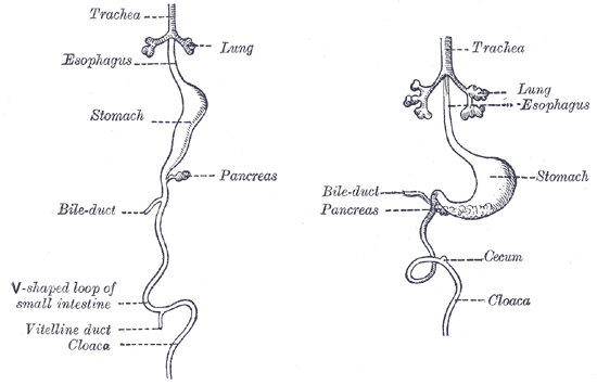 Front view of two successive stages in the development of the digestive tube.
