
Warthin's tumor biopsy
Editor-In-Chief: C. Michael Gibson, M.S., M.D. [1]; Associate Editor(s)-in-Chief: Ammu Susheela, M.D. [2]
Overview
Overview
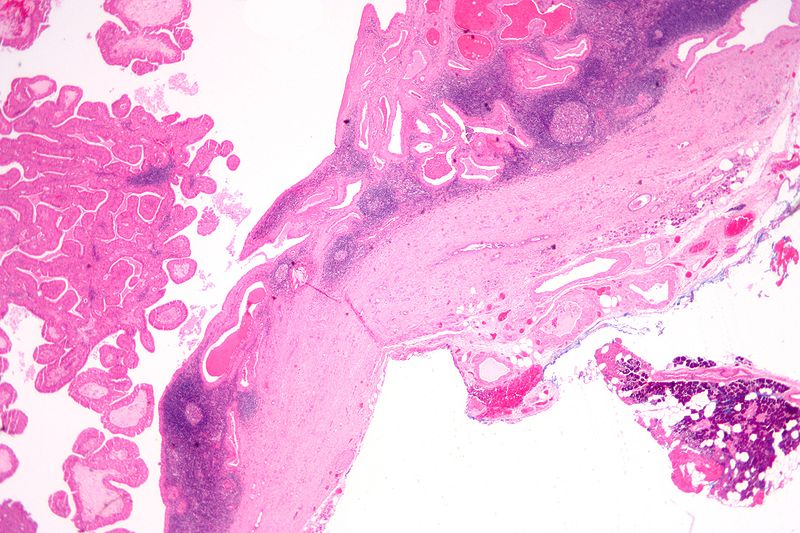
On biopsy, Warthin’s tumor is characterized by cystic spaces surrounded by two uniform rows of cells with centrally placed pyknotic nuclei, papillae with a two rows of pink epithelial cells, and lymphoid stroma.
Microscopic Pathology
Microscopic Pathology
- The appearance of this tumor under the microscope is unique. There are cystic spaces surrounded by two uniform rows of cells with centrally placed pyknotic nuclei.
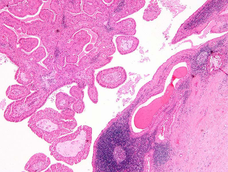
- The cystic spaces have epithelium referred to as papillary infoldings that protude into them. Additionally, the epithelium has lymphoid stroma with germinal center formation.
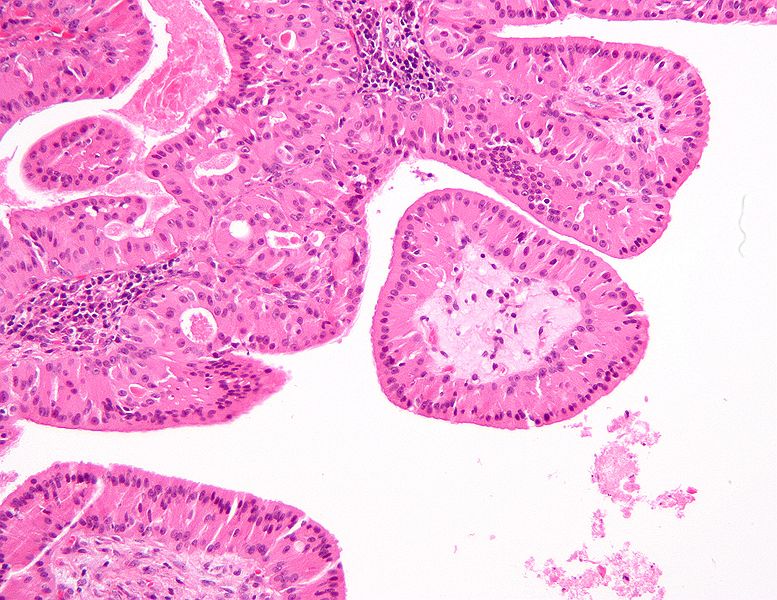
- Papillae (nipple-shaped structures) with a two rows of pink (eosinophilic) epithelial cells (with cuboidal basal cells and columnar luminal cells)
- Fibrous capsule – pink & homogenous on H&E stain
- Cystic space filled with debris in situ (not necrosis)
- Lymphoid stroma
- Additionally, the epithelium has lymphoid stroma with germinal center formation.[1]
-
![Histopathology of Warthin tumor in the parotid gland. H&E stain[2]](https://www.wikidoc.org/images/c/c7/Warthin_tumor_%281%29.jpg) Histopathology of Warthin tumor in the parotid gland. H&E stain[2]
Histopathology of Warthin tumor in the parotid gland. H&E stain[2] -
![Histopathology of Warthin tumor in the parotid gland. Another view of a file "Warthin tumor (1).jpg". H&E stain..[2]](https://www.wikidoc.org/images/c/cb/Warthin_tumor_%282%29.jpg) Histopathology of Warthin tumor in the parotid gland. Another view of a file “Warthin tumor (1).jpg”. H&E stain..[2]
Histopathology of Warthin tumor in the parotid gland. Another view of a file “Warthin tumor (1).jpg”. H&E stain..[2] -
![Histopathology of Warthin tumor in the parotid gland. Higher magnification of a file "Warthin tumor (1).jpg". H&E stain.[2]](https://www.wikidoc.org/images/d/d2/Warthin_tumor_%283%29.jpg) Histopathology of Warthin tumor in the parotid gland. Higher magnification of a file “Warthin tumor (1).jpg”. H&E stain.[2]
Histopathology of Warthin tumor in the parotid gland. Higher magnification of a file “Warthin tumor (1).jpg”. H&E stain.[2] -
 Histopathology of Warthin tumor in the parotid gland. Image courtesy of Nephron librepathology (original file ‘’here’’). Creative Commons BYSANC
 -
 Histopathology of Warthin tumor in the parotid gland. Image courtesy of Nephron librepathology (original file ‘’here’’). Creative Commons BYSANC
 -
Histopathology of Warthin tumor in the parotid gland. Image courtesy of Nephron librepathology (original file ‘’here’’). Creative Commons BYSANC
-
 Histopathology of Warthin tumor in the parotid gland. Image courtesy of Nephron librepathology (original file ‘’here’’). Creative Commons BYSANC
![Histopathology of Warthin tumor in the parotid gland. H&E stain[2]](https://www.wikidoc.org/index.php/File%3AWarthin_tumor_%281%29.jpg)
![Histopathology of Warthin tumor in the parotid gland. Another view of a file "Warthin tumor (1).jpg". H&E stain..[2]](https://www.wikidoc.org/index.php/File%3AWarthin_tumor_%282%29.jpg)
![Histopathology of Warthin tumor in the parotid gland. Higher magnification of a file "Warthin tumor (1).jpg". H&E stain.[2]](https://www.wikidoc.org/index.php/File%3AWarthin_tumor_%283%29.jpg)


References
References
- ↑ Warthin’s tumor librepathology (2015) http://librepathology.org/wiki/index.php/Warthin_tumour Accessed on December 14, 2015
- ↑ 2.0 2.1 2.2 Abid, Syed A.; Stack, Brendan C.; Bodenner, Donald L. (2014). “Metastatic Follicular Thyroid Carcinoma Secreting Thyroid Hormone and Radioiodine Avid without Stimulation: A Case Report and Literature Review”. Case Reports in Endocrinology. 2014: 1–6. doi:10.1155/2014/584513. ISSN 2090-6501.
Looking for the patient version?
© 2026 MyEClinic – IFTM Institut für Telematik in der Medizin GmbH