
Adenomyosis
For patient information, click here
Template:DiseaseDisorder infobox
Editor-In-Chief: C. Michael Gibson, M.S., M.D. [1]; Associate Editor(s)-In-Chief: Cafer Zorkun M.D. PhD.[2], Dina Elantably, MD[3]
Overview
Adenomyosis is a medical condition characterized by the presence of ectopic endometrial tissue (the inner lining of the uterus) within the myometrium (the thick, muscular layer of the uterus). The condition is typically found in women between the ages between 35 and 50. Patients with adenomyosis can have painful and/or profuse menses (dysmenorrhea & menorrhagia, respectively). Adenomyosis may involve the uterus focally, creating an adenomyoma, or diffusely. With diffuse involvement, the uterus becomes bulky and heavier.
Historical Perspective
- Adenomyosis was first discovered by Carl von Rokitansky, a German pathologist, in 1860 when he found endometrial glands in the myometrium and designated this finding as ‘cystosarcoma adenoids uterinum’[1].
- In 1892 the first systematic investigation of adenomyosis was carried out by ‘Thomas Stephen Cullen’, a gynecologist. He distinguished 3 types of adenomyoma: intramural, subperitoneal and submucous adenomyoma[1].
- In 1893, Kelly and Cullen described the pathogenesis of adenomyoma. The ‘gradual ascendancy of Cullen’s mucosal theory’ stated that endometrium invades the inner myometrium through the presence in it of ‘chinks’, or fissures.
- In 1892, Cullen described that abdominal hysterectomy is indicated for treatment as the endometrial growths are interwoven with the normal muscle of the uterus.
Historical Perspective
Discovery
- There is limited information about the historical perspective of [disease name].
OR
- [Disease name] was first discovered by [name of scientist], a [nationality + occupation], in [year]/during/following [event].
- The association between [important risk factor/cause] and [disease name] was made in/during [year/event].
- In [year], [scientist] was the first to discover the association between [risk factor] and the development of [disease name].
- In [year], [gene] mutations were first implicated in the pathogenesis of [disease name].
Landmark Events in the Development of Treatment Strategies
Impact on Cultural History
Famous Cases
The following are a few famous cases of [disease name]:
References
Classification
- Adenomyosis can be classified according to its histopathology into 2 groups:
- Diffuse adenomyosis: Uniformly enlarged boggy uterus.
- Focal adenomyosis (adenomyoma): Grossly it resembles fibroid but without a surrounding pseudocapsule.
- Other variants of adenomyosis include juvenile cystic adenomyosis; which is the presence of endometrial cysts > 1cm in diameter within the myometrium. It is usually seen in young women <30 years old [2].
Pathophysiology
- The pathogenesis of Adenomyosis is poorly understood. There two theories that explain the possible pathogenesis[3]:
- Endomyometrial invagination of the endometrium; due to weakness of the uterine smooth muscles.
- De novo development of adenomyosis from mullerian rests due to metaplasia.
- The basic Fibroblast Growth Factor (bFGF) receptor/ligand system has shown to be upregulated in adenomyosis which explain the abnormal uterine bleeding and menorrhagia[4].
- Estrogen and progesterone hormones play a role in the pathogenesis of adenomyosis[5]. Other hormones such as oxytocin [6], FSH[7], and prolactin[8] also contribute to the pathogenesis of the disease.
- On gross pathology, there is a globular enlargement of the myometrium of the uterus showing cysts filled with hemolysed red blood cells and sideroblasts[9].
- On microscopic histopathological analysis, there are endometrial glands, and stroma surrounded by hypertrophic smooth muscle bundles haphazardly scattered within the myometrium[9].
{{#ev:youtube|nOCtpIwCZ-Y}}
Pathophysiology is the study of the disturbance of normal mechanical, physical, and biochemical functions, either caused by a disease, or resulting from a disease or abnormal syndrome or condition that may not qualify to be called a disease.
An alternate definition is “the study of the biological and physical manifestations of disease as they correlate with the underlying abnormalities and physiological disturbances.”[1]
An example, from the field of infectious disease, would be the study of a toxin released by a bacterium, and what that toxin does to the body to cause harm, one possible result being sepsis. Another example is the study of the chemical changes that take place in body tissue due to inflammation.
Pathophysiology can be looked at as the intersection of two older, related disciplines: (normal) physiology and pathology.
Physiology is the study of normal, healthy bodily function (as opposed to anatomy, which is the study of normal structure). When something disrupts normal physiological processes, it enters the realm of pathophysiology.
Pathology, broadly speaking, is the “study of the nature and cause of disease.”[2] or the results of disease in the body. Pathophysiology looks at the detailed malfunctioning that comes from or, alternately, causes disease.
One caution in this approach is that healthy structure and function is not precisely the same in any two individuals.
Pathophysiology is a required study for under most nursing school programs in the United States as well as other countries.
See also
References
- Kumar, V., Abbas, A. and N. Fausto. 2004. Robbins & Cotran Pathologic Basis of Disease. Philadelphia: W. B. Saunders Company
Causes
The cause of adenomyosis is unknown, although it has been associated with any sort of uterine trauma that may break the barrier between the endometrium and myometrium, such as:
Differentiating adenomyosis from other Diseases
For further information about the differential diagnosis, click here.
Epidemiology and Demographics
- It is generally estimated that adenomyosis is present in 20-35% of women[10].
- The incidence and prevalence of adenomyosis are, however, difficult to be accurately estimated and biased by studying only women undergoing hysterectomy, so the total population of women having the disease is not known[11].
Age
- Adenomyosis is more commonly observed among women aged 40-50 years in those undergoing hysterectomy for diagnosis[9].
- Adenomyosis is less commonly diagnosed in adolescents who undergo pelvic imaging by transvaginal ultrasound or MRI rather than a hysterectomy for diagnosis[12].
Race
- There is no racial predilection for adenomyosis.
- Almost all cases of adenomyosis present in multiparous women, however there is no clear causal relationship between multiparty and the development of the disease[9]
Risk Factors
- Similar to the epidemiology, the risk factors of adenomyosis are unknown and difficult to be accurately determined as diagnosis is based on examining pathological specimens only in women undergoing hysterectomy[11].
- Adenomyosis often coexists with other pelvic diseases namely endometriosis and leiomyoma, so it is unknown whether it exhibits specific risk factors[12].
- Prior uterine surgery has been shown to be a possible risk factor for the development of adenomyosis[13].
Natural History, Complications and Prognosis
- Early clinical features of adenomyosis include dysmenorrhea, heavy menstrual bleeding, and chronic pelvic pain.
- Some reported complications of adenomyosis are preterm birth and miscarriage in young women diagnosed by pelvic imaging[14]. The relationship of adenomyosis to infertility is controversial[15].
- Prognosis is generally good as surgical treatment by hysterectomy is often curable unless there is another associated uterine pathology that requires further attention. There is no increased risk for secondary development of endometrial carcinoma.
Diagnosis
Adenomyosis is a histopathological diagnosis that is made after hysterectomy. The preoperative diagnosis is suggested by pelvic imaging such as transvaginal ultrasound and MRI along with the classical presentation of heavy menstrual bleeding, dysmenorrhea, and uniformly enlarged globular uterus.
- Symptoms of adenomyosis may include the following[16]:
- Heavy menstrual bleeding
- Dysmenorrhea
- Chronic pelvic pain
- Bimanual pelvic examination may be remarkable for[16]:
- Diffusely enlarged uterus (Boggy soft uterus)
- Uterus is mobile (not fixed as in endometriosis)
- Uterine tenderness may be noted.
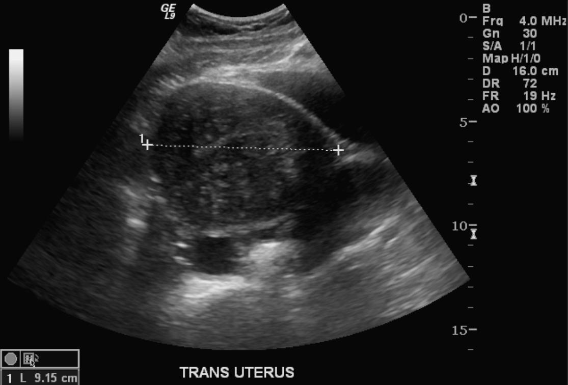
The uterus may be imaged using ultrasound (US) or magnetic resonance imaging (MRI). Transvaginal ultrasound is the most cost-effective and most available. Either modality will show an enlarged uterus. On ultrasound, the uterus will have a heterogeneous texture, without the focal well-defined masses that characterize uterine fibroids.
- Typical appearances of adenomyosis at transvaginal ultrasound include poorly marginated hypoechoic and heterogeneous areas within the myometrium, myometrial cysts, and a globular or enlarged uterus with asymmetry.
-
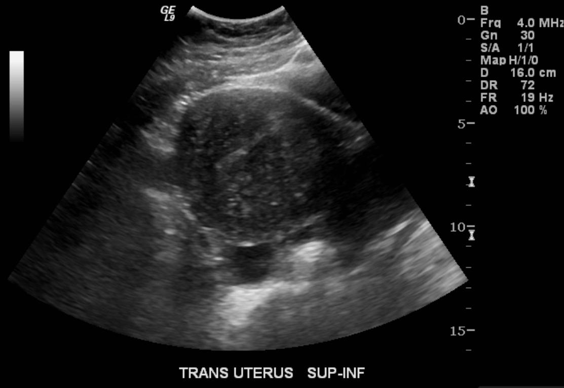 US: Adenomyosis
 -
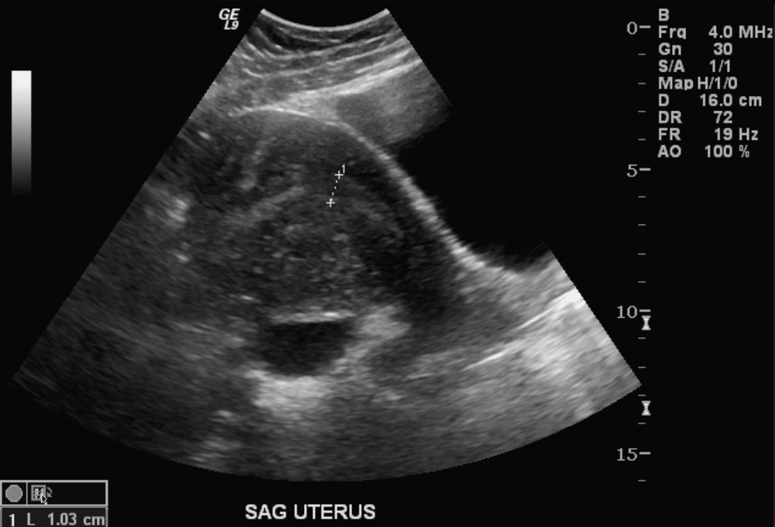 US: Adenomyosis
 -
 US: Adenomyosis


-
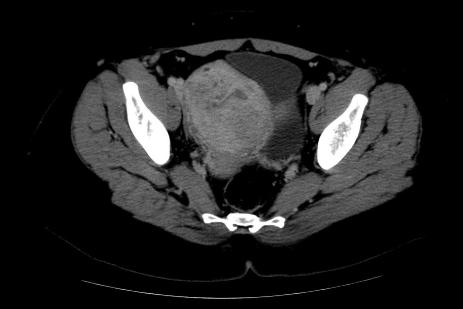 CT: Adenomyosis
 -
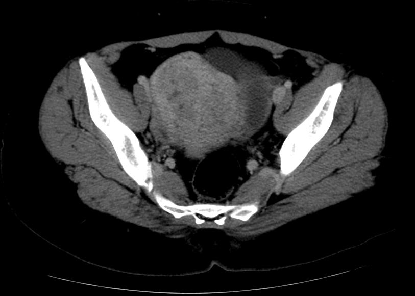 CT: Adenomyosis


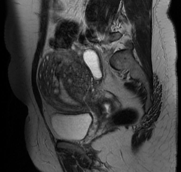
- MRI provides better diagnostic capability due to the increased spatial and contrast resolution, and to not be limited by the presence of bowel gas or calcified uterine fibroids (as is ultrasound). In particular, MRI is better able to differentiate adenomyosis from multiple small uterine fibroids.
- MRI can be used to classify adenomyosis based on the depth of penetration of the ectopic endometrium into the myometrium.
- Adenomyosis appears as either diffuse or focal thickening (greater than 12 mm )of the junctional zone forming an ill-defined area of low signal intensity, occasionally with embedded bright foci on T2-weighted images.
- Histologically, areas of low signal intensity correspond to smooth muscle hyperplasia, and bright foci on T2-weighted images correspond to islands of ectopic endometrial tissue and cystic dilatation of glands.
-
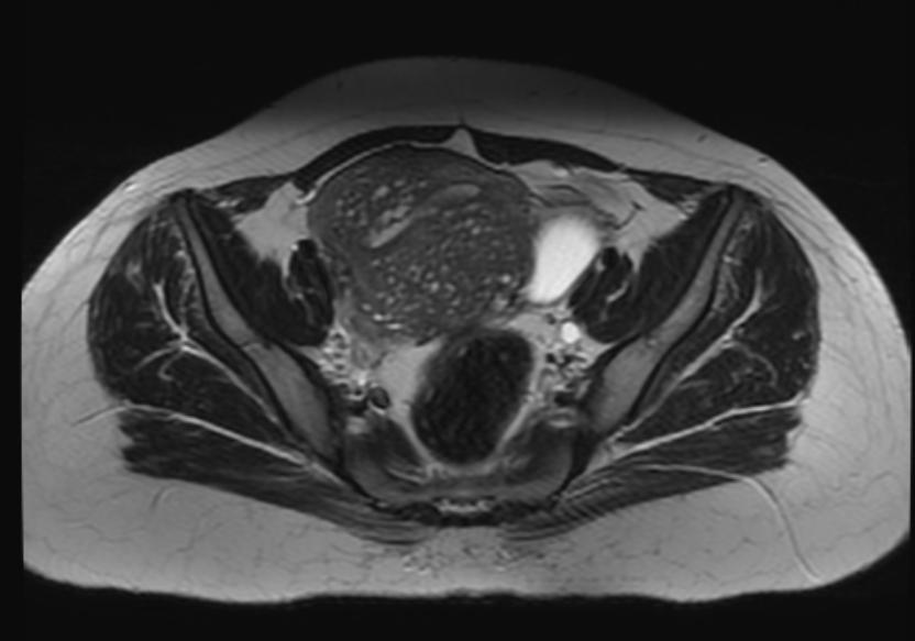 T2: Adenomyosis
 -
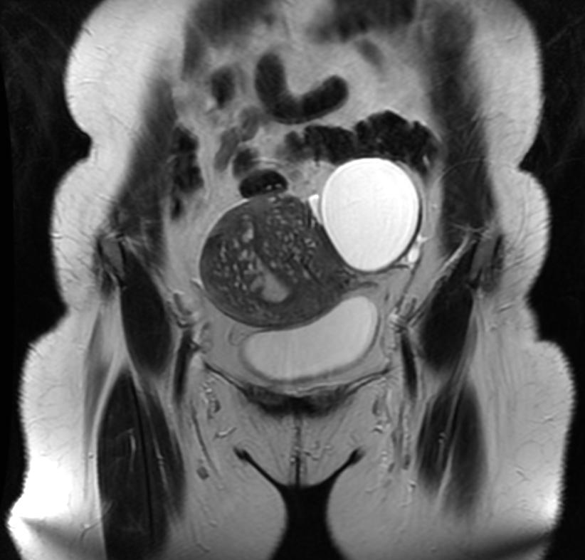 T2: Adenomyosis
 -
 T2: Adenomyosis
 -
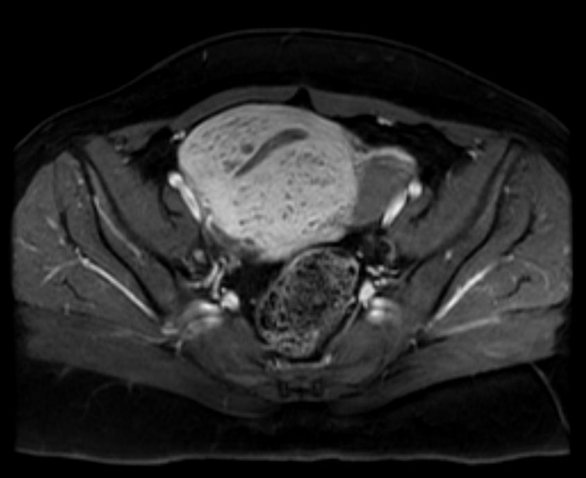 T1 fat sat contrast: Adenomyosis


Treatment
- Surgery is the mainstay of therapy for adenomyosis[17].
- Hysterectomy with preservation of the ovaries is the most common approach to the treatment of adenomyosis, and it is done via abdominal, transvaginal, laparoscopic approach, or robotic surgery. [17].
- Unlike Leiomyoma, there is no plane of cleavage to excise adenomyomas and preserve the uterus. Uterus sparing resection is an investigational approach especially for young women seeking future pregnancy[18].
- Medical treatment for dysmenorrhea and menorrhagia can be prescribed as a temporary alternative for young women in the child-bearing period.
- Hormonal therapy to control the symptoms includes levonorgestrel-releasing IUD (most preferred method), combined oral contraceptive pills, GnRH analogs, and oral GnRH antagonists[19].
- Levonorgestrel–IUD has a direct action on the uterus. It alleviates dysmenorrhea and menorrhagia[20].
- Once the hormonal therapy is stopped to conceive, symptoms recur within six months.
- In women who decline hysterectomy or have contraindications for hysterectomy or women who failed hormonal therapy, uterine artery embolization can be an alternative to control dysmenorrhea and heavy menstrual bleeding.[21].
- The outcomes of the procedure are significantly correlated with the lesion vascularity.[21].
Editor-In-Chief: C. Michael Gibson, M.S., M.D. [1]Sumanth Khadke, MD[2], Ogechukwu Hannah Nnabude, MD
Overview
Compliance with avoidance is important. The key to avoidance is proper evaluation and detection of causative allergen. Wear appropriate clothing to protect against irritants at home and in a work environment. [1] [2]
Treatment
High-potency topical corticosteroids, e.g. clobetasol propionate 0.05% cream, may be used to reduce the inflammation. [3] As a general rule, high-potency corticosteroids should not be used on thin skin, e.g. face, genitals, intertriginous areas, to avoid the risk of skin atrophy. Antihistamines such as hydroxyzine and cetirizine are recommended to control pruritus. Systemic steroids are advised in severe cases but should be tapered gradually to prevent recurrences. Friction should be avoided as well as the use of soaps, perfumes, and dyes. Emollients are used for hydrating the skin. Tacrolimus ointment and pimecrolimus cream are immunomodulating drugs that inhibit calcineurin and are helpful in allergic contact dermatitis.
Reference
- ↑ Soltanipoor M, Kezic S, Sluiter JK, de Wit F, Bosma AL, van Asperen R; et al. (2019). “Effectiveness of a skin care programme for the prevention of contact dermatitis in healthcare workers (the Healthy Hands Project): A single-centre, cluster randomized controlled trial”. Contact Dermatitis. 80 (6): 365–373. doi:10.1111/cod.13214. PMC 6593800 Check
|pmc=value (help). PMID 30652317. - ↑ Nedorost S (2018). “A diagnostic checklist for generalized dermatitis”. Clin Cosmet Investig Dermatol. 11: 545–549. doi:10.2147/CCID.S185357. PMC 6217130. PMID 30464569.
- ↑ Vernon HJ, Olsen EA (1990). “A controlled trial of clobetasol propionate ointment 0.05% in the treatment of experimentally induced Rhus dermatitis”. J Am Acad Dermatol. 23 (5 Pt 1): 829–32. doi:10.1016/0190-9622(90)70297-u. PMID 2147698.
References
- ↑ 1.0 1.1 Benagiano G, Brosens I (2006). “History of adenomyosis”. Best Pract Res Clin Obstet Gynaecol. 20 (4): 449–63. doi:10.1016/j.bpobgyn.2006.01.007. PMID 16515887.
- ↑ Takeuchi H, Kitade M, Kikuchi I, Kumakiri J, Kuroda K, Jinushi M (2010). “Diagnosis, laparoscopic management, and histopathologic findings of juvenile cystic adenomyoma: a review of nine cases”. Fertil Steril. 94 (3): 862–8. doi:10.1016/j.fertnstert.2009.05.010. PMID 19539912.
- ↑ Ferenczy A (1998). “Pathophysiology of adenomyosis”. Hum Reprod Update. 4 (4): 312–22. doi:10.1093/humupd/4.4.312. PMID 9825847.
- ↑ Propst AM, Quade BJ, Gargiulo AR, Nowak RA, Stewart EA (2001). “Adenomyosis demonstrates increased expression of the basic fibroblast growth factor receptor/ligand system compared with autologous endometrium”. Menopause. 8 (5): 368–71. doi:10.1097/00042192-200109000-00012. PMID 11528364.
- ↑ Green AR, Styles JA, Parrott EL, Gray D, Edwards RE, Smith AG; et al. (2005). “Neonatal tamoxifen treatment of mice leads to adenomyosis but not uterine cancer”. Exp Toxicol Pathol. 56 (4–5): 255–63. doi:10.1016/j.etp.2004.10.001. PMID 15816354.
- ↑ Guo SW, Mao X, Ma Q, Liu X (2013). “Dysmenorrhea and its severity are associated with increased uterine contractility and overexpression of oxytocin receptor (OTR) in women with symptomatic adenomyosis”. Fertil Steril. 99 (1): 231–240. doi:10.1016/j.fertnstert.2012.08.038. PMID 22999795.
- ↑ Stewart EA (2001). “Gonadotropins and the uterus: is there a gonad-independent pathway?”. J Soc Gynecol Investig. 8 (6): 319–26. PMID 11750866.
- ↑ Mori T, Singtripop T, Kawashima S (1991). “Animal model of uterine adenomyosis: is prolactin a potent inducer of adenomyosis in mice?”. Am J Obstet Gynecol. 165 (1): 232–4. doi:10.1016/0002-9378(91)90258-s. PMID 1853904.
- ↑ 9.0 9.1 9.2 9.3 Bergeron C, Amant F, Ferenczy A (2006). “Pathology and physiopathology of adenomyosis”. Best Pract Res Clin Obstet Gynaecol. 20 (4): 511–21. doi:10.1016/j.bpobgyn.2006.01.016. PMID 16563870.
- ↑ Vercellini P, Viganò P, Somigliana E, Daguati R, Abbiati A, Fedele L (2006). “Adenomyosis: epidemiological factors”. Best Pract Res Clin Obstet Gynaecol. 20 (4): 465–77. doi:10.1016/j.bpobgyn.2006.01.017. PMID 16563868.
- ↑ 11.0 11.1 Abbott JA (2017). “Adenomyosis and Abnormal Uterine Bleeding (AUB-A)-Pathogenesis, diagnosis, and management”. Best Pract Res Clin Obstet Gynaecol. 40: 68–81. doi:10.1016/j.bpobgyn.2016.09.006. PMID 27810281.
- ↑ 12.0 12.1 Parker JD, Leondires M, Sinaii N, Premkumar A, Nieman LK, Stratton P (2006). “Persistence of dysmenorrhea and nonmenstrual pain after optimal endometriosis surgery may indicate adenomyosis”. Fertil Steril. 86 (3): 711–5. doi:10.1016/j.fertnstert.2006.01.030. PMID 16782099.
- ↑ Panganamamula UR, Harmanli OH, Isik-Akbay EF, Grotegut CA, Dandolu V, Gaughan JP (2004). “Is prior uterine surgery a risk factor for adenomyosis?”. Obstet Gynecol. 104 (5 Pt 1): 1034–8. doi:10.1097/01.AOG.0000143264.59822.73. PMID 15516398.
- ↑ Horton J, Sterrenburg M, Lane S, Maheshwari A, Li TC, Cheong Y (2019). “Reproductive, obstetric, and perinatal outcomes of women with adenomyosis and endometriosis: a systematic review and meta-analysis”. Hum Reprod Update. 25 (5): 592–632. doi:10.1093/humupd/dmz012. PMID 31318420.
- ↑ Maheshwari A, Gurunath S, Fatima F, Bhattacharya S (2012). “Adenomyosis and subfertility: a systematic review of prevalence, diagnosis, treatment and fertility outcomes”. Hum Reprod Update. 18 (4): 374–92. doi:10.1093/humupd/dms006. PMID 22442261.
- ↑ 16.0 16.1 McElin TW, Bird CC (1974). “Adenomyosis of the uterus”. Obstet Gynecol Annu. 3 (0): 425–41. PMID 4608783.
- ↑ 17.0 17.1 Vannuccini S, Petraglia F (2019). “Recent advances in understanding and managing adenomyosis”. F1000Res. 8. doi:10.12688/f1000research.17242.1. PMC 6419978. PMID 30918629.
- ↑ Wood C (1998). “Surgical and medical treatment of adenomyosis”. Hum Reprod Update. 4 (4): 323–36. doi:10.1093/humupd/4.4.323. PMID 9825848.
- ↑ Schlaff WD, Ackerman RT, Al-Hendy A, Archer DF, Barnhart KT, Bradley LD; et al. (2020). “Elagolix for Heavy Menstrual Bleeding in Women with Uterine Fibroids”. N Engl J Med. 382 (4): 328–340. doi:10.1056/NEJMoa1904351. PMID 31971678.
- ↑ Bragheto AM, Caserta N, Bahamondes L, Petta CA (2007). “Effectiveness of the levonorgestrel-releasing intrauterine system in the treatment of adenomyosis diagnosed and monitored by magnetic resonance imaging”. Contraception. 76 (3): 195–9. doi:10.1016/j.contraception.2007.05.091. PMID 17707716.
- ↑ 21.0 21.1 Zhou J, He L, Liu P, Duan H, Zhang H, Li W; et al. (2016). “Outcomes in Adenomyosis Treated with Uterine Artery Embolization Are Associated with Lesion Vascularity: A Long-Term Follow-Up Study of 252 Cases”. PLoS One. 11 (11): e0165610. doi:10.1371/journal.pone.0165610. PMC 5091759. PMID 27806072.
Editor-In-Chief: C. Michael Gibson, M.S., M.D. [1]
Overview
WikiDoc has a reference manager that allows users to insert references based upon their PubMed ID number. If you move the text, the reference moves with it! All the references are automatically inserted at the bottom. You click on the reference and you go to the article. It is simple!
Where do I type in the references?
Usually you would type a number in the text like this (1) or this 1 which refers to a reference you would like to cite. You would then type in the reference at the end of the article. This is not the preferred method in WikiDoc.
WikiDoc features an automated reference manager. The advantages of this reference manager are that:
- The references are numbered automatically!
- When you move the text, the reference moves with it!
- You can click on the reference and go directly to the article!
Unlike the usual format where you type in the references at the end of the article, when you use the WikiDoc reference manager, you insert code for the reference right after the material you want to add a reference to. You use a software program to add in the references. The next section describes how to use the software program.
Using the WikiDoc reference manager
One goal of WikiDoc is to create reference lists that allow you to click on the PubMed ID number at the end of the reference and go to the primary article itself. This greatly facilitates locating references. Therefore, the preferred method of citing references includes the use of a reference manager that uses the PubMed ID number to create the reference. The reference manager uses not only PubMed IDs but also DrugBank ID, HGNC ID, ISBN, PubMed ID, PubMed Central ID, PubChem ID, or URLs.
The preferred method for inserting a reference is as follows:
Step 1: Use PubMed to locate the article you are interested in by clicking here
Step 2: Copy the PubMed ID number from the article. The word PubMed ID is often abbreviated PMID and this number is located at the bottom of the abstract. You can use the search function on Firefox or Internet explorer to locate the word PMID and the number will follow this abbreviation. It will look something like this:
PMID: 19032997
Step 3: Paste the PubMed ID number (PMID) into a software program that creates the Wiki language code for the reference you are going to paste into the text of your article. You can access software for converting a pubmed ID number from one of the following:
- SUMSearch Biomedical Citation Maker (converts PMID into wiki reference markups with embedded sumsearch link)
- DOI Wikipedia reference generator (converts DOI into wiki reference markups)
- DTU Informatics PMID to Cite journal (converts PMID into wiki reference markups)
- OttoBib (converts ISBN into wiki reference markups)
- Dave’s Template Filler (converts DrugBank ID, HGNC ID, ISBN, PubMed ID, PubMed Central ID, PubChem ID, or URL into wiki reference markups)
Step 4: When using the reference manager, make sure the button that says add ref tag is checked
Step 5: Press submit to generate the Wiki language that can be inserted in your article.
Step 6: Copy the Wiki language from the software program. For example, the output for the PMID above (19032997) is as follows:
<ref name=”pmid19032997″>{{cite journal |author=Gibson CM, Pride YB, Frederick PD, ”et al.” |title=Trends in reperfusion strategies, door-to-needle and door-to-balloon times, and in-hospital mortality among patients with ST-segment elevation myocardial infarction enrolled in the National Registry of Myocardial Infarction from 1990 to 2006 |journal=Am. Heart J. |volume=156 |issue=6 |pages=1035–44 |year=2008 |month=December |pmid=19032997 |doi=10.1016/j.ahj.2008.07.029 |url=}}</ref>
Step 7: Paste this output from the reference manager software right where you want the superscript number to appear in the text.
For example, using the above reference, you might type something like this:
Gibson et al recently summarized the improvements made in clincial outcomes as a result of 15 years of quality improvement efforts in the NRMI registry <ref name=”pmid19032997″>{{cite journal |author=Gibson CM, Pride YB, Frederick PD, ”et al.” |title=Trends in reperfusion strategies, door-to-needle and door-to-balloon times, and in-hospital mortality among patients with ST-segment elevation myocardial infarction enrolled in the National Registry of Myocardial Infarction from 1990 to 2006 |journal=Am. Heart J. |volume=156 |issue=6 |pages=1035–44 |year=2008 |month=December |pmid=19032997 |doi=10.1016/j.ahj.2008.07.029 |url=}}</ref>
This would generate text that looks like this:
Gibson et al recently summarized the improvements made in clincial outcomes as a result of 15 years of quality improvement efforts in the NRMI registry[1]
- ↑ Gibson CM, Pride YB, Frederick PD; et al. (2008). “Trends in reperfusion strategies, door-to-needle and door-to-balloon times, and in-hospital mortality among patients with ST-segment elevation myocardial infarction enrolled in the National Registry of Myocardial Infarction from 1990 to 2006”. Am. Heart J. 156 (6): 1035–44. doi:10.1016/j.ahj.2008.07.029. PMID 19032997. Unknown parameter
|month=ignored (help)
What if I don’t want to use the reference manager? What if I just want to type the references in myself?
You are not required to use the reference manager. You can do the following:
Step 1: Place a <ref> … </ref> immediately after the sentence where you want a footnote number to appear.
Step 2: Type the text of the note between the ref tags. For example if I typed this
This was on of the first articles I published <ref>J Fam Pract. 2000 Oct;49(10):921-3.</ref>
It would appear as this
This was one of the first articles I published[1]
Note: If you intend on using the same manual reference more than once, you will need to assign a name to the reference. You can do this by writing <ref name=”anythingyouwanthere”> … </ref>. Your name can be anything you want. Without a name, every manual reference will be viewed as a unique reference. This means that a reference used multiple times will show up as multiple references instead of just the one.
How do I make the references appear at the bottom of the article?
The preferred method
Put the following code in:
==References==
{{Reflist|2}}
This will generate your references in small font, in two columns, with links to the original article and abstract. It will return text that looks like this:
- ↑ J Fam Pract. 2000 Oct;49(10):921-3.
Additional Resources
- Atri M, Reinhold C, Mehio AR. Adenomyosis: US features with histologic correlation in an in-vitro study. Radiology. Jun 2000;215(3):783-90.
- Batzer FR, Hansen L. Bizarre sonographic appearance of an adenomyoma and its presentation. J Ultrasound Med. Aug 1996;15(8):599-602.
- Bazot M, Daraï E. [Evaluation of pelvic endometriosis: the role of MRI.]. J Radiol. Nov 2008;89(11 Pt 1):1695-6.
- Byun JY, Kim SE, Choi BG, et al. Diffuse and focal adenomyosis: MR imaging findings. Radiographics. Oct 1999;19 Spec No:S161-70.
- Chiang CH, Chang MY, Hsu JJ. Tumor vascular pattern and blood flow impedance in the differential diagnosis of leiomyoma and adenomyosis by color Doppler sonography. J Assist Reprod Genet. May 1999;16(5):268-75.
- Guilbeault H, Wilson SR, Lickrish GM. Massive uterine enlargement with necrosis: an unusual manifestation of adenomyosis. J Ultrasound Med. Apr 1994;13(4):326-8.
- Haimovici JB, Tempany CM. MR of the female pelvis: benign disease. Appl Radiol. Jun 1994;7:21.
- Huang HY. Medical treatment of endometriosis. Chang Gung Med J. Sep-Oct 2008;31(5):431-40.
- Iribarne C, Plaza J, De la Fuente P, et al. Intramyometrial cystic adenomyosis. J Clin Ultrasound. Jun 1994;22(5):348-50.
- Jarlot C, Anglade E, Paillocher N, Moreau D, Catala L, Aubé C. [MR imaging features of deep pelvic endometriosis: correlation with laparoscopy.]. J Radiol. Nov 2008;89(11 Pt 1):1745-54.
- Kang S, Turner DA, Foster GS, et al. Adenomyosis: specificity of 5 mm as the maximum normal uterine junctional zone thickness in MR images. AJR Am J Roentgenol. May 1996;166(5):1145-50.
- Kim MD, Lee HS, Lee MH, Kim HJ, Cho JH, Cha SH. Long-term results of symptomatic fibroids treated with uterine artery embolization: In conjunction with MR evaluation. Eur J Radiol. Dec 10 2008;
- Ostrzenski A. Extensive iatrogenic adenomyosis after laparoscopic myomectomy. Fertil Steril. Jan 1998;69(1):143-5.
- Piketty M, Chopin N, Dousset B, Millischer-Bellaische AE, Roseau G, Leconte M, et al. Preoperative work-up for patients with deeply infiltrating endometriosis: transvaginal ultrasonography must definitely be the first-line imaging examination. Hum Reprod. Dec 17 2008;
- Piketty M, Chopin N, Dousset B, Millischer-Bellaische AE, Roseau G, Leconte M, et al. Preoperative work-up for patients with deeply infiltrating endometriosis: transvaginal ultrasonography must definitely be the first-line imaging examination. Hum Reprod. Dec 17 2008;
- Reinhold C, Atri M, Mehio A, et al. Diffuse uterine adenomyosis: morphologic criteria and diagnostic accuracy of endovaginal sonography. Radiology. Dec 1995;197(3):609-14.
- Reinhold C, Tafazoli F, Mehio A, et al. Uterine adenomyosis: endovaginal US and MR imaging features with histopathologic correlation. Radiographics. Oct 1999;19 Spec No:S147-60.
- Tamai K, Koyama T, Umeoka S, et al. Spectrum of MR features in adenomyosis. Best Pract Res Clin Obstet Gynaecol. Aug 2006;20(4):583-602.
- Tamai K, Togashi K, Ito T, et al. MR imaging findings of adenomyosis: correlation with histopathologic features and diagnostic pitfalls. Radiographics. Jan-Feb 2005;25(1):21-40.
- Utsunomiya D, Notsute S, Hayashida Y, et al. Endometrial carcinoma in adenomyosis: assessment of myometrial invasion on T2-weighted spin-echo and gadolinium-enhanced T1-weighted images. AJR Am J Roentgenol. Feb 2004;182(2):399-404.
Template:Diseases of the pelvis, genitals and breasts
Looking for the patient version?
© 2026 MyEClinic – IFTM Institut für Telematik in der Medizin GmbH