
Angiogram


Editor-In-Chief: C. Michael Gibson, M.S., M.D. [1]
Overview
Overview
Angiography or arteriography is a medical imaging technique in which an X-ray picture is taken to visualize the inner opening (lumen) of blood filled structures, including arteries, veins and the heart chambers. Its name comes from the Greek words angeion, “vessel”, and graphien, “to write or record”. The X-ray film or image of the blood vessels is called an angiograph, or more commonly, an angiogram.
Historical Aspect
Historical Aspect
The term angiography, or angeiography, was originally used of a description of the weights, measures, vessels, etc, used by several nations.
The Portuguese physician and neurologist Egas Moniz, Nobel Prize winner in 1949, developed in 1927 the technique of contrasted x-ray cerebral angiography to diagnose several kinds of nervous diseases, such as tumors and arteriovenous malformations.
He is usually recognized as one of the pioneers in this field.
With the introduction of the Seldinger technique in 1953, the procedure became markedly safer as no sharp introductory devices needed to remain inside the vascular lumen.
Methods
Methods
Angiograms require the insertion of a catheter into a peripheral artery, e.g. the femoral artery.
As blood has the same radiodensity as the surrounding tissues, a radiocontrast agent (which absorbs X-rays) is added to the blood to make angiography visualization possible. The angiographic X-ray image shows shadows of the openings within the cardiovascular structures carrying blood (actually the radiocontrast agent within). The blood vessels or heart chambers themselves remain largely to totally invisible on the X-ray image.
The X-ray images may be taken as either still images, displayed on a fluoroscope or film, useful for mapping an area. Alternatively, they may be motion images, usually taken at 30 frames per second, which also show the speed of blood (actually the speed of radiocontrast within the blood) traveling within the blood vessel.
The most common angiogram performed is to visualize the blood in the coronary arteries. A long, thin, flexible tube called a catheter is used so as to administer the radiocontrast agent at the desired area to be visualized. The catheter is threaded into an artery in the groin or forearm, and the tip is advanced through the arterial system into one of the two major coronary arteries. X-ray images of the transient radiocontrast distribution within the blood flowing within the coronary arteries allows visualization of the size of the artery openings. Presence or absence of atherosclerosis or atheroma within the walls of the arteries cannot be clearly determined. See coronary catheterization for more detail.
Angiography is also commonly performed to identify vessel narrowing in patients with retinal vascular disorders, such as diabetic retinopathy and macular degeneration.
Clinical Applications
Clinical Applications
Coronary Angiography
One of most common angiograms performed is to visualize the blood in the coronary arteries. A long, thin, flexible tube called a catheter is used to administer the x-ray contrast agent at the desired area to be visualized. The catheter is threaded into an artery in the groin or forearm, and the tip is advanced through the arterial system into one of the two major coronary arteries.
X-ray images of the transient radiocontrast distribution within the blood flowing within the coronary arteries allows visualization of the size of the artery openings. Presence or absence of atherosclerosis or atheroma within the walls of the arteries cannot be clearly determined. See coronary catheterization for more detail..
Neuro-vascular angiography
Another increasingly common angiographic procedure is neuro-vascular digital subtraction angiography in order to visualise the arterial and venous supply to the brain. Intervention work such as coil-embolisation of aneurysms and AVM gluing can also be performed
Peripheral Angiography
Angiography is also commonly performed to identify vessel narrowing in patients with leg claudication or cramps, caused by reduced blood flow down the legs and to the feet; in patients with renal stenosis (which commonly causes high blood pressure) and can be used in the head to find and repair stroke. These are all done routinely through the femoral artery, but can also be performed through the brachial or axillary (arm) artery. Any stenoses found may be treated by the use of angioplasty.
Other
Other angiographic uses include the diagnosis of retinal vascular disorders, such as diabetic retinopathy and macular degeneration.
Types of angiographs
Types of angiographs
- Cerebral angiography
- Coronary angiography
- Peripheral angiography (arm or leg)
- Visceral angiography (the abdominal organs, or viscera)
- Pulmonary angiography (lungs)
- Lymphangiography (lymph vessels)
- Right heart ventriculography (looking at the right side of the heart)
- Left heart ventriculography (looking at the left side of the heart)
- Aortography (looking at the aorta, the major artery from the heart)
- Retinal angiography
- Magnetic Resonance Angiography

Postmortem Angiograms: Coronary Arteries
Postmortem Angiograms: Coronary Arteries
-
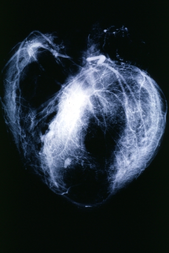
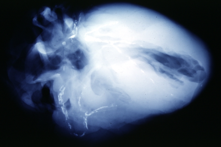
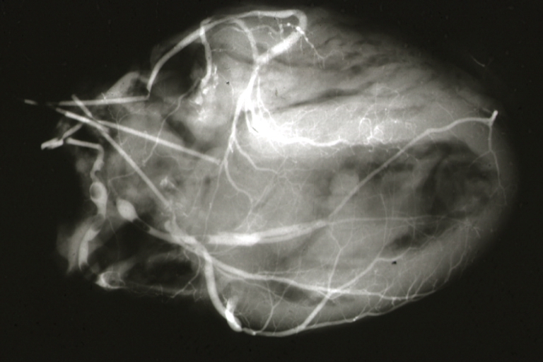 Myocardial Infarction: Postmortem angiogram of coronary arteries
 -
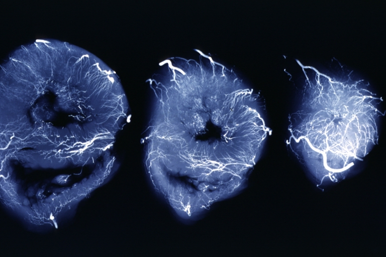
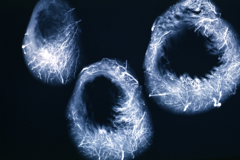 Angiogram: X-ray, horizontal sections of ventricle showing penetrating artery distribution (a quite good example)
 -
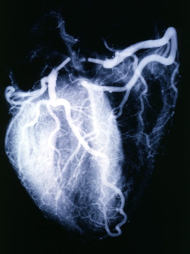
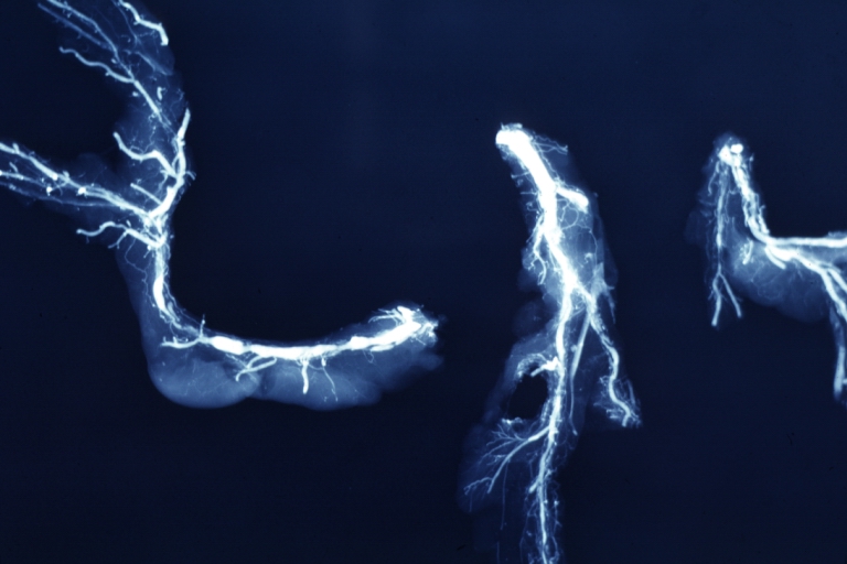
 Angiogram: X-ray, postmortem coronary arteries with multiple lesions
 -
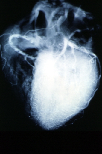
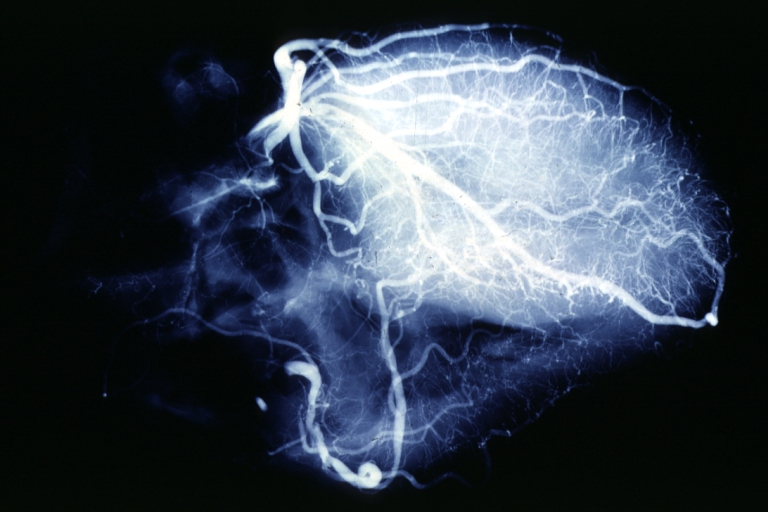
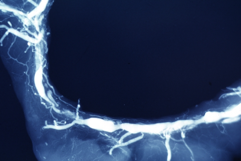
 Angiogram: X-ray postmortem normal coronaries
 -
 Angiogram: Postmortem angiogram with apparent lesions in proximal right coronary artery
 -
 Angiogram: X-ray, postmortem injection horizontal slice of left ventricle showing very well penetrating arteries
 -
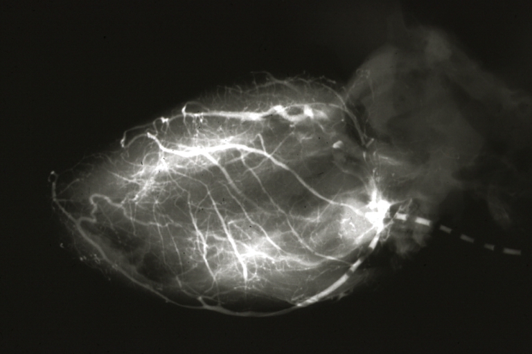
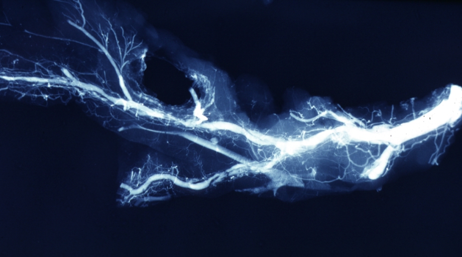
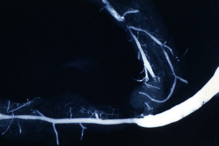 Angiogram Saphenous Vein Bypass Graft: X-ray shows rather close-up large vein anastomosing to much smaller artery
 -
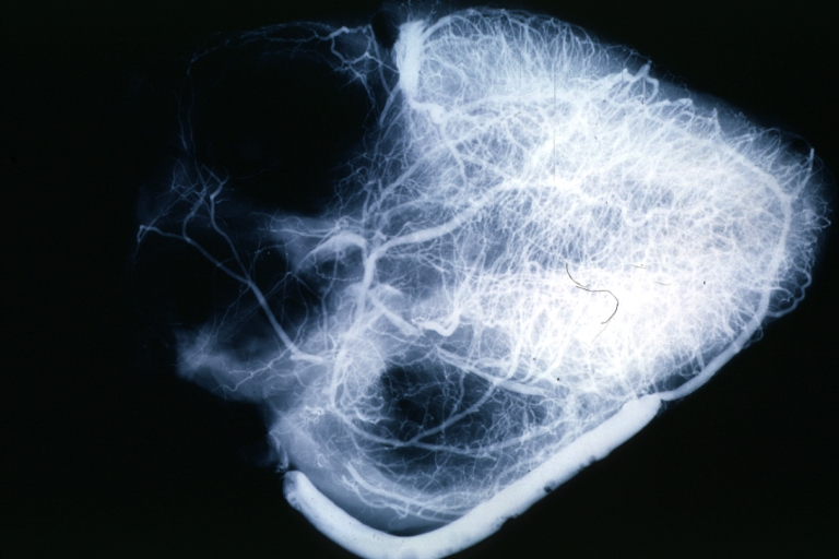 Angiogram Saphenous Vein Bypass Graft: X-ray postmortem injection showing vein anastomosis very well and the vasculature of the right and left ventricles
 -
 Coronary artery: Atherosclerosis: X-ray postmortem extensive lesions in this x-ray of whole heart
 -
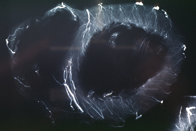
 X-Ray Intramyocardial Arteries: X-ray three horizontal slices of ventricles showing quite well the penetrating arteries
 -
 X-Ray Intramyocardial Arteries: X-ray three horizontal slices of ventricles showing quite well the penetrating arteries
 -
 Coronary Artery Anomalous Origin; Left From Pulmonary Artery: Angiogram, postmortem, after switch of left coronary artery to aorta
 -
 Coronary artery: Atherosclerosis: X-ray, postmortem, dissected arteries and extensive lesions
 -
 Coronary artery: Atherosclerosis: X-ray, postmortem, close-up view of artery with extensive lesions (very good example)
 -
 Coronary artery: Atherosclerosis: X-ray, postmortem, dissected artery, lesions in small branches


External links
External links
- RadiologyInfo – The radiology information resource for patients: Angiography procedures
- Cardiac Catheterization from Angioplasty.Org
- Angiography Equipment from Siemens Medical
- Cardiovascular and Interventional Radiological Society of Europe
- Worldwide Angiography Manufacturer
cs:Angiografie de:Angiografie it:Angiografia nl:Angiografie no:Angiografi fi:Angiografia
Looking for the patient version?
© 2026 MyEClinic – IFTM Institut für Telematik in der Medizin GmbH