
Atrial septal defect chest x ray
Editor-In-Chief: C. Michael Gibson, M.S., M.D. [1]; Associate Editor(s)-In-Chief: Priyamvada Singh, M.B.B.S. [2]; Cafer Zorkun, M.D., Ph.D. [3]; Assistant Editor(s)-In-Chief: Kristin Feeney, B.S. [4]
Overview
Overview
Chest x rays may detect an atrial septal defect. Chest x rays can be limited in imaging quality and may only supplement other imaging modalities. The chest x-ray may demonstrate cardiomegaly (right ventricle and right atrial enlargement), a prominent pulmonary artery segment and increased pulmonary vascular markings.
Chest X Ray
Chest X Ray
Common Findings
CXR findings on an anteroposterior view of the chest x-ray in atrial septal defect may include: [1]
1) Prominent pulmonary artery, increased pulmonary vascular markings.
2) Cardiomegaly due to right atrial and ventricular enlargement.
3)’ Triangular appearance of the heart
- Results from enlargement of pulmonary arteries preventing the ascending and transverse aorta from forming normal heart borders.
- A vertical, modestly curved, density in the right-side of the pericardium, may be visible.
- Commonly associated with the sinus venosus atrial septal defect.
- Results from the point of insertion of the pulmonary vein into the inferior vena cava.
- May cause abnormal densities within the chest x ray.
5) Dilatation of the superior vena cava can be seen in sinus venosus
Less Common Findings
- Normal appearance of heart vasculature
- Left heart enlargement/left atrial enlargement
- Pulmonary edema
- Pulmonary venous hypertension
Imagings
-
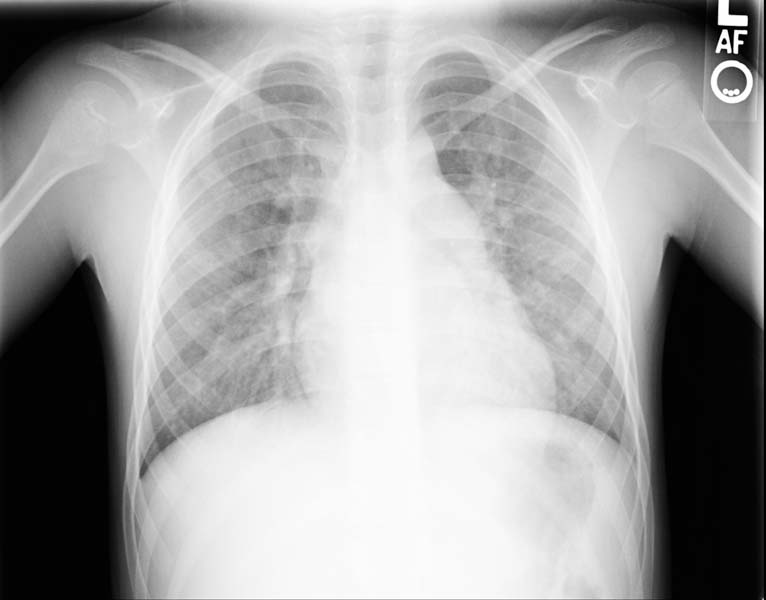 Enlarged right atrial border and mild cardiomegaly.
 -
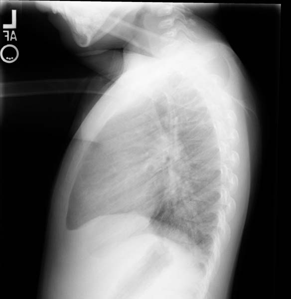 Lateral view
 -
 Post repair. Enlarged right atrial border and mild cardiomegaly.
 -
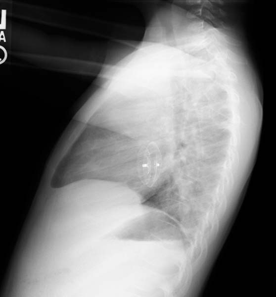 Post repair. Lateral view.
 -
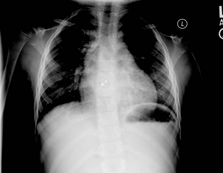
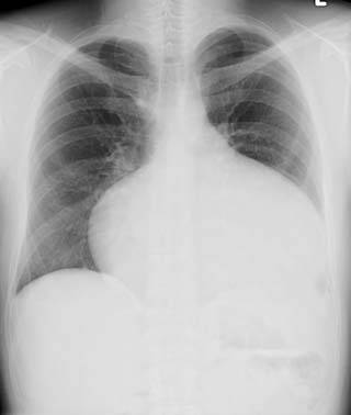 ASD. Another patient. Enlarged right atrial border and advanced cardiomegaly.


References
References
- ↑ Abdulla, Ra-id. (2011). Heart Diseases in Children: A Pediatrician’s Guide. Springer.
Looking for the patient version?
© 2026 MyEClinic – IFTM Institut für Telematik in der Medizin GmbH