
Left atrial enlargement
Editor-In-Chief: C. Michael Gibson, M.S., M.D. [1]
Causes of Left atrial enlargement
Editor-In-Chief: C. Michael Gibson, M.S., M.D. [4]; Associate Editor(s)-In-Chief: Varun Kumar, M.B.B.S. [5]
Overview
In the general population, obesity appears to be the most important risk factor for left atrial enlargement.[1] Persistent atrial fibrillation results in retention of blood in the left atrium secondary to inadequate pumping of blood by the atrium into the left ventricle[2]. Similar pathophysiologic process occurs in mitral stenosis resulting in subsequent left atrial enlargement. Aortic stenosis, mitral regurgitation, aortic insufficiency and other causes of left ventricular heart failure causes an increase in back pressure within the atrium resulting in subsequent left atrial enlargement.
Differential Diagnosis of Causes of Left Atrial Enlargement
By Organ System
| Cardiovascular | Aortic regurgitation, Aortic stenosis, Atrial fibrillation[2], Atrial septal defect, Cardiac amyloidosis[3], Coarctation of the aorta, Congestive heart failure, Coronary artery disease, Hypertrophic cardiomyopathy, Hypoplasia of mitral valve annulus which is a part of the spectrum of hypoplastic left heart syndrome, Infective endocarditis, Ischemic mitral regurgitation, Left atrial myxoma, Left bundle branch block, Mitral regurgitation, Mitral stenosis, Mitral valve annular calcification, Mitral valve commissural fusion, Mitral valve prolapse, Papillary muscle rupture, Parachute mitral valve, Prosthetic mitral valve stenosis, Rheumatic heart disease, Supra-valve mitral membrane, Systemic hypertension, Thickened chordae tendinae, Thrombus (ball valve thrombus) in left atrium |
| Chemical / poisoning | No underlying causes |
| Dermatologic | No underlying causes |
| Drug Side Effect | Chronic alcoholism[4] |
| Ear Nose Throat | No underlying causes |
| Endocrine | No underlying causes |
| Environmental | No underlying causes |
| Gastroenterologic | No underlying causes |
| Genetic | Cardiac amyloidosis[3], Hypertrophic cardiomyopathy, Hypoplasia of mitral valve annulus which is a part of the spectrum of hypoplastic left heart syndrome |
| Hematologic | No underlying causes |
| Iatrogenic | No underlying causes |
| Infectious Disease | Infective endocarditis, Pneumonia, Rheumatic heart disease |
| Musculoskeletal / Ortho | Ankylosing spondylitis |
| Neurologic | No underlying causes |
| Nutritional / Metabolic | Hypercholesterolemia accelerates the stenotic process of the valves via calcification, Obesity |
| Obstetric/Gynecologic | No underlying causes |
| Oncologic | Left atrial myxoma |
| Opthalmologic | No underlying causes |
| Overdose / Toxicity | Chronic alcoholism[5] |
| Psychiatric | No underlying causes |
| Pulmonary | Emphysema, Pneumonia |
| Renal / Electrolyte | No underlying causes |
| Rheum / Immune / Allergy | Ankylosing spondylitis, Cardiac amyloidosis[3], Rheumatic heart disease |
| Sexual | No underlying causes |
| Trauma | No underlying causes |
| Urologic | No underlying causes |
| Miscellaneous | Thrombus (ball valve thrombus) in left atrium |
In Alphabetical Order
- Ankylosing spondylitis
- Aortic regurgitation
- Aortic stenosis
- Atrial fibrillation[2]
- Atrial septal defect
- Cardiac amyloidosis[3]
- Chronic alcoholism[6]
- Coarctation of the aorta
- Congestive heart failure
- Coronary artery disease
- Emphysema
- Hypertrophic cardiomyopathy
- Hypercholesterolemia accelerates the stenotic process of the valves via calcification
- Hypoplasia of mitral valve annulus which is a part of the spectrum of hypoplastic left heart syndrome
- Infective endocarditis
- Ischemic mitral regurgitation
- Left atrial myxoma
- Left bundle branch block
- Mitral regurgitation
- Mitral stenosis
- Mitral valve annular calcification
- Mitral valve commissural fusion
- Mitral valve prolapse
- Obesity
- Papillary muscle rupture
- Parachute mitral valve
- Pneumonia
- Prosthetic mitral valve stenosis
- Rheumatic heart disease
- Supra-valve mitral membrane
- Systemic hypertension
- Thickened chordae tendinae
- Thrombus (ball valve thrombus) in left atrium
References
- ↑ Stritzke J, Markus MRP, Duderstadt S, Lieb W, Luchner A, Döring A, Keil U, Hense H and Schunkert H (2009-11-17). “The Aging Process of the Heart: Obesity Is the Main Risk Factor for Left Atrial Enlargement During Aging: The MONICA/KORA (Monitoring of Trends and Determinations in Cardiovascular Disease/Cooperative Research in the Region of Augsburg) Study”. Journal of the American College of Cardiology. 54 (21): 1982–9. doi:10.1016/j.jacc.2009.07.034. PMID 19909880. Retrieved 2009-12-02.
- ↑ 2.0 2.1 2.2 Sanfilippo AJ, Abascal VM, Sheehan M, Oertel LB, Harrigan P, Hughes RA and Weyman AE (1990). “Atrial enlargement as a consequence of atrial fibrillation A prospective echocardiographic study”. Circulation. 82 (3): 792–7. doi:10.1161/01.CIR.82.3.792. PMID 2144217. Retrieved 2009-12-02.
- ↑ 3.0 3.1 3.2 3.3 Dardas PS, Tsikaderis DD, Mezilis N, Geleris P, Boudoulas H (2002). “Echocardiographic evidence of atrial myopathy in amyloidosis: a case report”. European Journal of Echocardiography : the Journal of the Working Group on Echocardiography of the European Society of Cardiology. 3 (4): 303–5. PMID 12413446. Retrieved 2012-04-17. Unknown parameter
|month=ignored (help) - ↑ Kabir J. Singh, Beth Cohen, Bee Ya Na, Mary A. Whooley, and Nelson B. Schiller. Predictors Of Increasing Left Atrial Volume Over Five Years In Coronary Artery Disease Patients: Results From The Heart And Soul Study. J.Am.Coll.Cardiol.2010;55;A91.E864 [1]
- ↑ Kabir J. Singh, Beth Cohen, Bee Ya Na, Mary A. Whooley, and Nelson B. Schiller. Predictors Of Increasing Left Atrial Volume Over Five Years In Coronary Artery Disease Patients: Results From The Heart And Soul Study. J.Am.Coll.Cardiol.2010;55;A91.E864 [2]
- ↑ Kabir J. Singh, Beth Cohen, Bee Ya Na, Mary A. Whooley, and Nelson B. Schiller. Predictors Of Increasing Left Atrial Volume Over Five Years In Coronary Artery Disease Patients: Results From The Heart And Soul Study. J.Am.Coll.Cardiol.2010;55;A91.E864 [3]
Electrocardiogram Findings
Editor-In-Chief: C. Michael Gibson, M.S., M.D. [2]; Associate Editor-In-Chief: Cafer Zorkun, M.D., Ph.D. [3]
Overview
Left atrial enlargement may be observed among patients with pressure or volume overload of the left atrium.
Differential Diagnosis of Left Atrial Enlargement
- Atrial aneurysm
- Infective endocarditis
- Left heart failure
- Mitral regurgitation
- Mitral stenosis
- Mitral valve prolapse
- Myxedema
- Patent Ductus Arteriosus
- Ventricular septal defect
EKG in Left Atrial Enlargement
Left atrial enlargement produces a broad, bifid P wave in lead II (P mitrale) and enhances the terminal negative portion (negative deflection) of the P wave in V1.
In lead I and/or II the following may be seen:

In lead V1, the following may be seen:
- Biphasic P wave with terminal negative portion > 40 ms duration (>0.04 sec or 1 small square)
- Biphasic P wave with terminal negative portion > 1mm deep

Examples of Left Atrial Enlargement on EKG
-
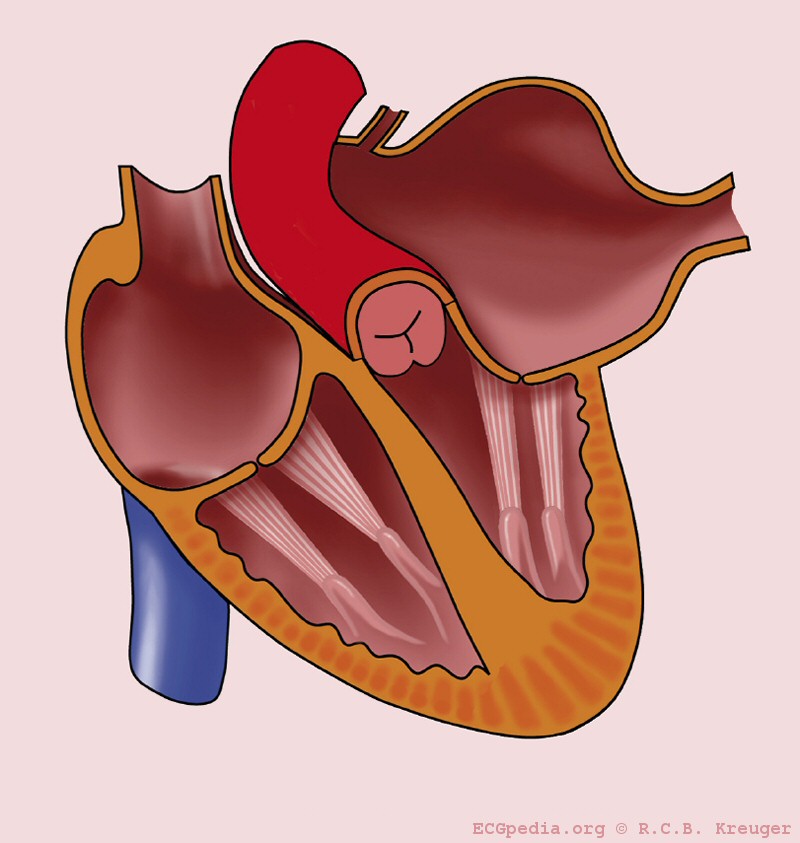 Left atrial enlargement
 -
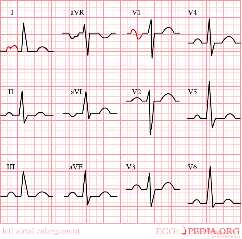 12 lead EKG: Left atrial enlargement


-
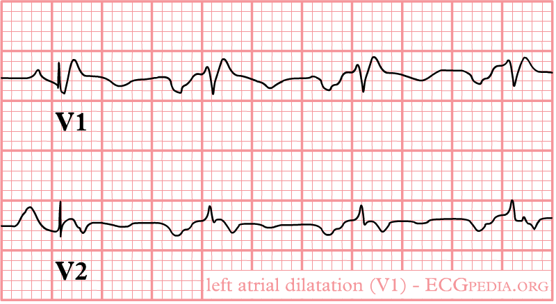 Left atrial enlargement as seen in lead V1.
 -
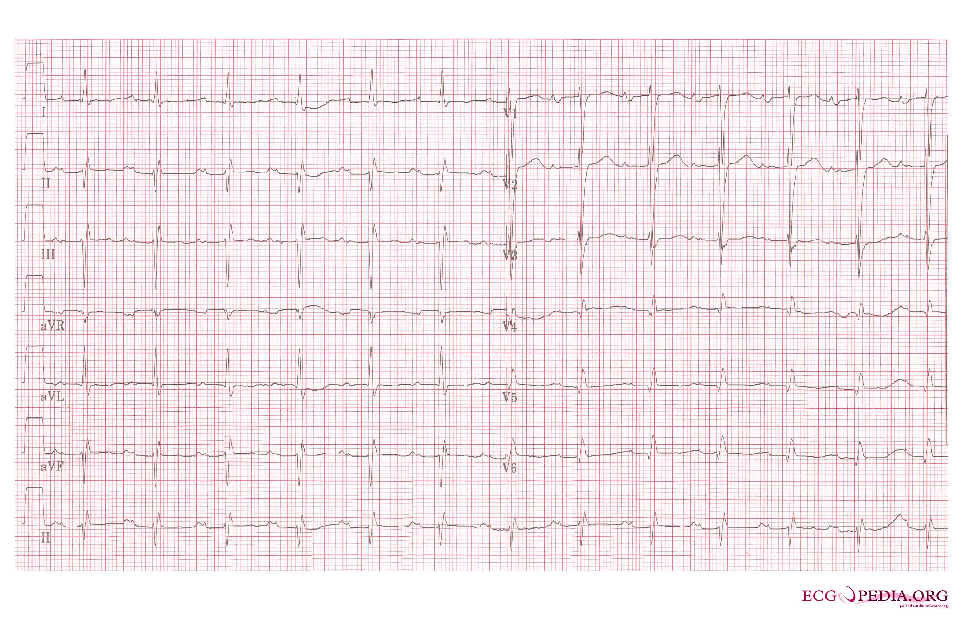 Left atrial enlargement, a 12 lead ECG

See Also
Chest X-Ray Findings
Editor-In-Chief: C. Michael Gibson, M.S., M.D. [1]; Associate Editor(s)-In-Chief: Cafer Zorkun, M.D., Ph.D. [2]; Varun Kumar, M.B.B.S. [3]
Chest X-Ray
Chest x-ray findings of left atrial enlargement are:
- Double density sign: Occur when the right side of the left atrium pushes behind the right atrial border, appearing as a double density. If large enough it can actually reach beyond the border of the right atrium.
- Convex left atria appendage: usually reflect prior rheumatic heart disease
- Splaying of the carina
- Posterior displacement of the left main stem bronchus on lateral radiograph
- Superior displacement of the left main stem bronchus on frontal view
- Posterior displacement of a barium filled oesophagus or nasogastric tube
Images shown below are courtesy of Radiopedia.com.
-
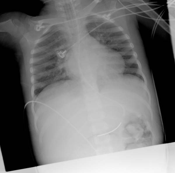 Double density sign
 -
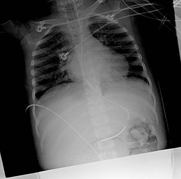 Same patient & the same image. Double density sign. Image’s modified for more contrast and better visualization.
 -
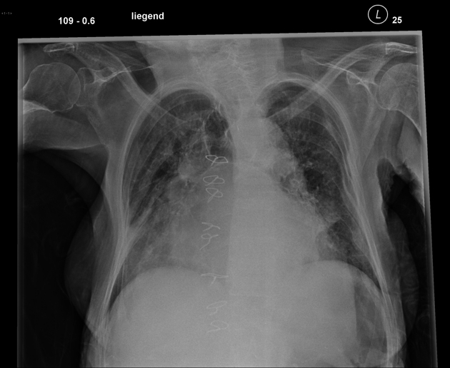 Aside from the dirty lung due to emphysema and pneumonic infiltration in the lower right field you can notice a marked enlargement of the left atrium with splaying of the carina.


References
Looking for the patient version?
© 2026 MyEClinic – IFTM Institut für Telematik in der Medizin GmbH