

Health Dictionary
Reliable, easy-to-read patient information on common medical conditions — written in plain language, free of jargon, and grounded in current evidence. Browse the most frequent diagnoses, understand what they mean for everyday life, and learn which steps and questions help you have a focused conversation with your doctor.
Atrial septal defect chest x ray
Introduction
Overview
Overview
Chest x rays may detect an atrial septal defect. Chest x rays can be limited in imaging quality and may only supplement other imaging modalities. The chest x-ray may demonstrate cardiomegaly (right ventricle and right atrial enlargement), a prominent pulmonary artery segment and increased pulmonary vascular markings.
Chest X Ray
Chest X Ray
Common Findings
CXR findings on an anteroposterior view of the chest x-ray in atrial septal defect may include: [1]
1) Prominent pulmonary artery, increased pulmonary vascular markings.
2) Cardiomegaly due to right atrial and ventricular enlargement.
3)’ Triangular appearance of the heart
- Results from enlargement of pulmonary arteries preventing the ascending and transverse aorta from forming normal heart borders.
- A vertical, modestly curved, density in the right-side of the pericardium, may be visible.
- Commonly associated with the sinus venosus atrial septal defect.
- Results from the point of insertion of the pulmonary vein into the inferior vena cava.
- May cause abnormal densities within the chest x ray.
5) Dilatation of the superior vena cava can be seen in sinus venosus
Less Common Findings
- Normal appearance of heart vasculature
- Left heart enlargement/left atrial enlargement
- Pulmonary edema
- Pulmonary venous hypertension
Imagings
-
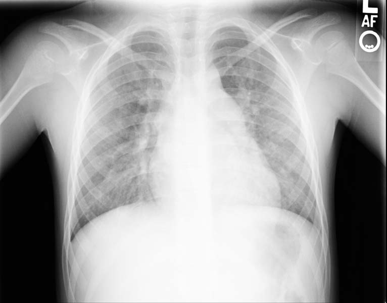 Enlarged right atrial border and mild cardiomegaly.
 -
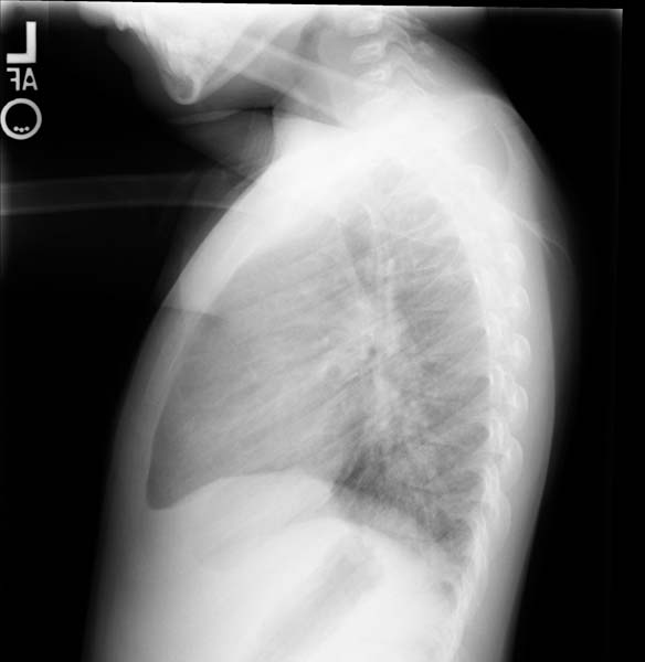 Lateral view
 -
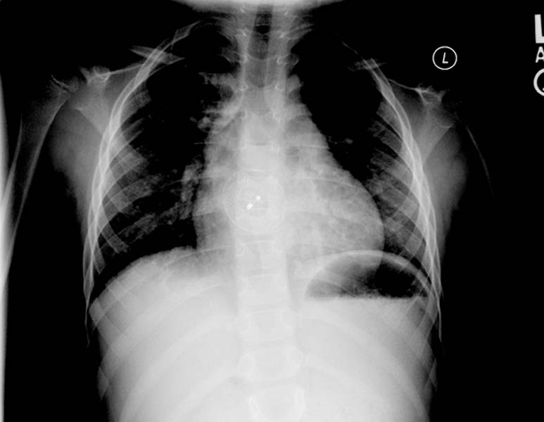 Post repair. Enlarged right atrial border and mild cardiomegaly.
 -
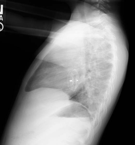 Post repair. Lateral view.
 -
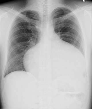 ASD. Another patient. Enlarged right atrial border and advanced cardiomegaly.


References
References
- ↑ Abdulla, Ra-id. (2011). Heart Diseases in Children: A Pediatrician’s Guide. Springer.
Editor-In-Chief: C. Michael Gibson, M.S., M.D. [1]; Associate Editor(s)-In-Chief: Priyamvada Singh, M.B.B.S. [2]; Cafer Zorkun, M.D., Ph.D. [3]; Assistant Editor(s)-In-Chief: Kristin Feeney, B.S. [4]