

Health Dictionary
Reliable, easy-to-read patient information on common medical conditions — written in plain language, free of jargon, and grounded in current evidence. Browse the most frequent diagnoses, understand what they mean for everyday life, and learn which steps and questions help you have a focused conversation with your doctor.
Blepharitis pathophysiology
Introduction
Overview
Overview
The exact pathogenesis of blepharitis is not fully understood. It is thought that blepharitis is caused by either bacterial colonization in the eyelids or meibomian gland dysfunction. Blepharitis may also be caused by allergens and mites that affect the eyelashes.[1][2]
Pathophysiology
Pathophysiology
Pathogenesis
The exact pathogenesis of blepharitis is not fully understood. It is thought that blepharitis is caused by either bacterial colonization in the eyelids or meibomian gland dysfunction. However, in most cases of blepharitis, there is a considerable pathogenesis overlap between anterior and posterior blepharitis.
Anterior blepharitis
Anterior blepharitis is often associated with staphylococcal infection or seborrhoeic dermatitis. The exact pathogenesis of anterior blepharitis is not fully understood. It is thought that blepharitis is caused by bacterial colonization (Staphylococcus aureus or Staphylococcus epidermidis) in the eyelids. Following bacterial colonization, bacterial lipase changes meibomian gland secretions and increases cholesterol concentration. These changes may result in an environment that affects the ocular surface and tear evaporation. It is thought that anterior blepharitis may also be caused by allergic response to bacterial antigens (mostly staphylococcal antigens).[1][2]
Posterior blepharitis
The exact pathogenesis of posterior blepharitis is not fully understood. It is thought that the abnormal meibomian gland secretions may result in posterior blepharitis. Posterior blepharitis is often associated with skin conditions, such as rosacea or seborrhoeic dermatitis. These conditions are associated with sebaceous glands and gland orifices abnormalities. It is thought that the abnormal meibomian gland secretions may cause a direct toxic effect on the ocular surface. Sebaceous glands abnormalities may also result in an environment that affect bacterial growth.[3]
Acute blepharitis
Acute blepharitis may be caused by irritant or allergen exposure.[4]
Chronic blepharitis
Demodex folliculorum is an external parasite in hair follicles, sebaceous glands, and meibomian glands. It may cause either anterior or posterior blepharitis. The exact pathogenesis of chronic blepharitis caused by the Demodex mites is not fully understood. It is thought that over-proliferation of Demodex folliculorum in meibomian glands may result in lid-margin infection and ocular discomfort. It is also thought demodex blepharitis may be caused by immune system reactions to the mites leading to inflammation. [5][6]
Associated Conditions
Many diseases are associated with blepharitis such as skin, systemic, and ocular disease. These include:
- Systemic or skin disease:[7][8]
- Ocular disease:[7][8][9]
- Conjunctivitis
- Meibomianitis (inflammation of the meibomian glands)
- Keratitis
- Dry eye syndrome
- Chalazion
- Trichiasis
Gross Pathology
On gross pathology, lid margin swelling, misdirection of lashes, loss of lashes, oily or greasy deposits and crusting of anterior lid margin, , and conjunctival hyperaemia are characteristic findings of blepharitis.[10]
Microscopic Pathology
On microscopic histopathological analysis, non granulomatous inflammation with neutrophils, acanthosis, or parakeratosis are characteristic findings of staphylococcal blepharitis.[11]
On microscopic histopathological analysis, hyperkeratinization of the meibomian gland ductal epithelium, mononuclear cellular infiltrates, and spongiosis in eyelids are characteristic findings of seborrheic blepharitis.[12]
On microscopic histopathological analysis, chronic inflammatory changes, epithelial hyperplasia, and follicular plugging are characteristic findings of chronic blepharitis.[13]
Images
The following are gross images associated with blepharitis:
-
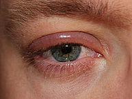 Blepharitis of the right eye – By clubtable – Own work (Original text: eigenes Foto), Public Domain, https://commons.wikimedia.org/w/index.php?curid=20138102
 -
 Infant with blepharitis – By Sage Ross – Own work, CC BY-SA 3.0, https://commons.wikimedia.org/w/index.php?curid=8978907


References
References
- ↑ 1.0 1.1 Bunya VY, Brainard DH, Daniel E, Massaro-Giordano M, Nyberg W, Windsor EA; et al. (2013). “Assessment of signs of anterior blepharitis using standardized color photographs”. Cornea. 32 (11): 1475–82. doi:10.1097/ICO.0b013e3182a02e0e. PMC 3947496. PMID 24055901.
- ↑ 2.0 2.1 Dougherty JM, McCulley JP (1986). “Bacterial lipases and chronic blepharitis”. Invest Ophthalmol Vis Sci. 27 (4): 486–91. PMID 3957566.
- ↑ Blepharitis. American Academy of Ophthalmology/eyewiki (2014). http://eyewiki.aao.org/Blepharitis Accessed on July 14, 2016
- ↑ Lemp MA, Nichols KK (2009). “Blepharitis in the United States 2009: a survey-based perspective on prevalence and treatment”. Ocul Surf. 7 (2 Suppl): S1–S14. PMID 19383269.
- ↑ Bhandari V, Reddy JK (2014). “Blepharitis: always remember demodex”. Middle East Afr J Ophthalmol. 21 (4): 317–20. doi:10.4103/0974-9233.142268. PMC 4219223. PMID 25371637.
- ↑ Viswalingam M, Rauz S, Morlet N, Dart JK (2005). “Blepharokeratoconjunctivitis in children: diagnosis and treatment”. Br J Ophthalmol. 89 (4): 400–3. doi:10.1136/bjo.2004.052134. PMC 1772603. PMID 15774912.
- ↑ 7.0 7.1 McCulley JP, Dougherty JM (1985). “Blepharitis associated with acne rosacea and seborrheic dermatitis”. Int Ophthalmol Clin. 25 (1): 159–72. PMID 3156100.
- ↑ 8.0 8.1 Nemet AY, Vinker S, Kaiserman I (2011). “Associated morbidity of blepharitis”. Ophthalmology. 118 (6): 1062–8. doi:10.1016/j.ophtha.2010.10.015. PMID 21276617.
- ↑ Blepharitis. Diseases Database (2016). http://www.diseasesdatabase.com/relationship.asp?glngUserChoice=1455&bytRel=2&blnBW=0&strBB=RL&blnClassSort=255 Accessed on July 15, 2016
- ↑ Benitez-Del-Castillo JM (2012). “How to promote and preserve eyelid health”. Clin Ophthalmol. 6: 1689–98. doi:10.2147/OPTH.S33133. PMC 3484726. PMID 23118519.
- ↑ Seal D, Ficker L, Ramakrishnan M, Wright P (1990). “Role of staphylococcal toxin production in blepharitis”. Ophthalmology. 97 (12): 1684–8. PMID 2087299.
- ↑ THYGESON P, VAUGHAN DG (1954). “Seborrheic blepharitis”. Trans Am Ophthalmol Soc. 52: 173–88. PMC 1312591. PMID 13274422.
- ↑ Wesolowska M, Knysz B, Reich A, Blazejewska D, Czarnecki M, Gladysz A; et al. (2014). “Prevalence of Demodex spp. in eyelash follicles in different populations”. Arch Med Sci. 10 (2): 319–24. doi:10.5114/aoms.2014.42585. PMC 4042053. PMID 24904668.
Editor-In-Chief: C. Michael Gibson, M.S., M.D. [1]; Associate Editor(s)-in-Chief: Sara Mehrsefat, M.D. [2]