
Calvaria (skull)
Editor-In-Chief: C. Michael Gibson, M.S., M.D. [1]
The calvaria (or calva, or skullcap) is the roof of the skull. It is formed by the following bones:
- frontal bone
- parietal bones (two)
- temporal bones (two)
- occipital bone
In a fetus, the formation of the Calvaria involves a process known as intramembranous ossification, although the base of the skull (underlying the brain) develops through endochondral ossification.
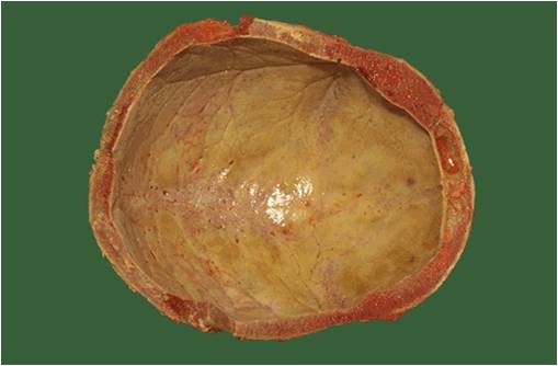
Inner surface of the skull-cap
Inner surface of the skull-cap
The inner surface of the skull-cap is concave and presents depressions for the convolutions of the cerebrum, together with numerous furrows for the lodgment of branches of the meningeal vessels.
Along the middle line is a longitudinal groove, narrow in front, where it commences at the frontal crest, but broader behind; it lodges the superior sagittal sinus, and its margins afford attachment to the falx cerebri.
On either side of it are several depressions for the arachnoid granulations, and at its back part, the openings of the parietal foramina when these are present.
It is crossed, in front, by the coronal suture, and behind by the lambdoidal, while the sagittal lies in the medial plane between the parietal bones.
(Images courtesy of Melih Aktan M.D. Istanbul Medical Faculty, Turkey)
-
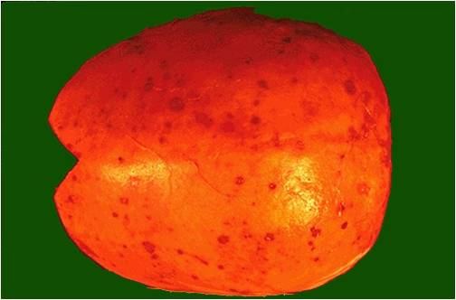 Calvarium in multiple myeloma
 -
 Calvarium in Thalassemia


External links
External links
References
References
- Tubbs, R Shane (2008). “The intriguing history of the human calvaria: sinister and religious”. Child’s nervous system : ChNS : official journal of the International Society for Pediatric Neurosurgery. Germany. 24 (4): 417–22. doi:10.1007/s00381-007-0509-0. ISSN 0256-7040. PMID 18026961. Unknown parameter
|month=ignored (help); Unknown parameter|quotes=ignored (help); Unknown parameter|coauthors=ignored (help)
Looking for the patient version?
© 2026 MyEClinic – IFTM Institut für Telematik in der Medizin GmbH