
Cavernous sinus
Editor-In-Chief: C. Michael Gibson, M.S., M.D. [1]
The cavernous sinus (or lateral sellar compartment) is a large collection of thin-walled veins creating a cavity bordered by the sphenoid bone and the temporal bone of the skull.
Contents
Contents
Each cavernous sinus (one for each hemisphere of the brain) contains the following:
- vertically, from superior to inferior
- oculomotor nerve (CN III)
- trochlear nerve (CN IV)
- ophthalmic nerve, the V1 branch of the trigeminal nerve (CN V)
- maxillary nerve, the V2 branch of CN V
- horizontally
- internal carotid artery (and sympathetic plexus). See also cavernous part of internal carotid artery.
- abducens nerve (CN VI)
One mnemonic for remembering the contents is “OTOM CAT”[1]
Venous connections
Venous connections
It receives tributaries from:
- Superior and inferior ophthalmic veins
- Superior parietal sinus
- Superior and middle cerebral veins
The veins of exit are to the superior and inferior petrosal sinuses as well as via the emissary veins through the foramina of the skull. There are also connections with the pterygoid plexus of veins via inferior ophthalmic vein, deep facial vein and emissary veins.
Clinical significance
Clinical significance
It is the only anatomic location in the body in which an artery travels completely through a venous structure. If the internal carotid artery ruptures within the cavernous sinus, an arteriovenous fistula is created (more specifically, a carotid-cavernous fistula).
The pituitary gland lies directly below the cavernous sinus. An abnormally growing pituitary adenoma, surrounded on all other sides by the bony walls of the sella turcica, will expand in the direction of least resistance and eventually compress the cavernous sinus. Cavernous sinus syndrome may result from mass effect of these tumors and cause ophthalmoplegia (from compression of the oculomotor nerve, trochlear nerve, and abducens nerve), ophthalmic sensory loss (from compression of the ophthalmic nerve), and maxillary sensory loss (from compression of the maxillary nerve).
Additional images
Additional images
-
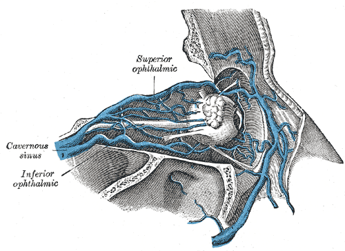 Veins of orbit.
 -
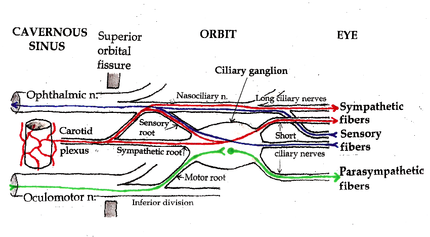 Pathways in the Ciliary Ganglion.


External links
External links
- Template:LoyolaMedEd
- Template:SUNYAnatomyFigs – “Venous dural sinuses.”
- Cavernous+Sinus at the US National Library of Medicine Medical Subject Headings (MeSH)
- Template:NormanAnatomy
- UMichAtlas|n3a8p1
- Template:EMedicineDictionary
Looking for the patient version?
© 2026 MyEClinic – IFTM Institut für Telematik in der Medizin GmbH