
Chickenpox physical examination
Editor-In-Chief: C. Michael Gibson, M.S., M.D. [1];Associate Editor(s)-in-Chief: Aravind Reddy Kothagadi M.B.B.S[2]
Overview
Overview
The diagnosis of varicella is primarily clinical. Skin lesions on physical examination include pruritic macules on the back, chest, face, abdomen and extremities. Skin lesions progress to papules and heal by crusting. The other common skin lesions include papules, vesicles, pustules and crusts.
Physical Examination
Physical Examination
Appearance of the Patient
- The patient appears weak and tired with rashes spread over the scalp, face, trunk, and limbs. The intense pruritis triggers recurrent urges to scratch.
Vitals
- Low-grade fever
- Tachycardia[1]
Skin
Skin lesions on physical examination include:
- Pruritic macules on the back, chest, face, abdomen and extremities.
- Skin lesions progress to papules and heal by crusting. Other common skin lesions include:
HEENT
Lungs
Normal breath sounds are heard.
Abdomen
The abdomen will not be tender and there is no organomegaly.
Heart
- Normal S1 and S2 are heard.
- Ventricular fibrillation presumed to be secondary to myocarditis has been observed in some of the adult population. [1]
CNS
- The neurological examination may be normal with no focal neurological deficits.
- CNS may show the following findings:
- Meningitis
- Meningoencephalitis
- Vasculopathy
Gallery
Gallery
-
![Chickenpox lesions on the skin of this patient's left breast and arm on day 6 of the illness. From Public Health Image Library (PHIL). [2]](https://www.wikidoc.org/images/9/95/Chickenpox36.jpeg) Chickenpox lesions on the skin of this patient’s left breast and arm on day 6 of the illness. From Public Health Image Library (PHIL). [2]
Chickenpox lesions on the skin of this patient’s left breast and arm on day 6 of the illness. From Public Health Image Library (PHIL). [2] -
![Chickenpox lesions on the skin of this patient's back and buttocks at day 6 of the illness. From Public Health Image Library (PHIL). [2]](https://www.wikidoc.org/images/8/8e/Chickenpox35.jpeg) Chickenpox lesions on the skin of this patient’s back and buttocks at day 6 of the illness. From Public Health Image Library (PHIL). [2]
Chickenpox lesions on the skin of this patient’s back and buttocks at day 6 of the illness. From Public Health Image Library (PHIL). [2] -
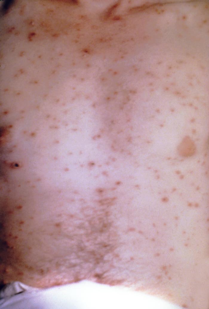
![Chickenpox lesions on the skin of this patient's breasts, arms, and torso at day 6 of the illness. From Public Health Image Library (PHIL). [2]](https://www.wikidoc.org/images/7/71/Chickenpox34.jpeg) Chickenpox lesions on the skin of this patient’s breasts, arms, and torso at day 6 of the illness. From Public Health Image Library (PHIL). [2]
Chickenpox lesions on the skin of this patient’s breasts, arms, and torso at day 6 of the illness. From Public Health Image Library (PHIL). [2] -
![Patient with cervical skin lesions caused by chickenpox. From Public Health Image Library (PHIL). [2]](https://www.wikidoc.org/images/c/c7/Chickenpox33.jpeg) Patient with cervical skin lesions caused by chickenpox. From Public Health Image Library (PHIL). [2]
Patient with cervical skin lesions caused by chickenpox. From Public Health Image Library (PHIL). [2] -
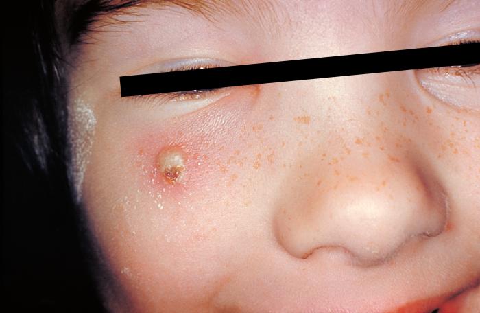
![4-month old infant with skin lesions on his brow ridge due to chickenpox. From Public Health Image Library (PHIL). [2]](https://www.wikidoc.org/images/0/03/Chickenpox32.jpeg) 4-month old infant with skin lesions on his brow ridge due to chickenpox. From Public Health Image Library (PHIL). [2]
4-month old infant with skin lesions on his brow ridge due to chickenpox. From Public Health Image Library (PHIL). [2] -
![Patient had presented with chickenpox demonstrating the typical rash on day eight. From Public Health Image Library (PHIL). [2]](https://www.wikidoc.org/images/8/84/Chickenpox29.jpeg) Patient had presented with chickenpox demonstrating the typical rash on day eight. From Public Health Image Library (PHIL). [2]
Patient had presented with chickenpox demonstrating the typical rash on day eight. From Public Health Image Library (PHIL). [2] -
![Patient developed palatal mucosal lesions due to chickenpox. From Public Health Image Library (PHIL). [2]](https://www.wikidoc.org/images/1/18/Chickenpox28.jpeg) Patient developed palatal mucosal lesions due to chickenpox. From Public Health Image Library (PHIL). [2]
Patient developed palatal mucosal lesions due to chickenpox. From Public Health Image Library (PHIL). [2] -
![Vaccine recipient developed a secondary herpes infection adjacent to the vaccination site. From Public Health Image Library (PHIL). [2]](https://www.wikidoc.org/images/3/3e/Chickenpox27.jpeg) Vaccine recipient developed a secondary herpes infection adjacent to the vaccination site. From Public Health Image Library (PHIL). [2]
Vaccine recipient developed a secondary herpes infection adjacent to the vaccination site. From Public Health Image Library (PHIL). [2] -
![Pustulovesicular rash represents a generalized herpes outbreak due to the Varicella-zoster virus (VZV) pathogen. From Public Health Image Library (PHIL). [2]](https://www.wikidoc.org/images/6/6e/Chickenpox26.jpeg) Pustulovesicular rash represents a generalized herpes outbreak due to the Varicella-zoster virus (VZV) pathogen. From Public Health Image Library (PHIL). [2]
Pustulovesicular rash represents a generalized herpes outbreak due to the Varicella-zoster virus (VZV) pathogen. From Public Health Image Library (PHIL). [2] -
![Case of chickenpox. From Public Health Image Library (PHIL). [2]](https://www.wikidoc.org/images/e/e2/Chickenpox24.jpeg) Case of chickenpox. From Public Health Image Library (PHIL). [2]
Case of chickenpox. From Public Health Image Library (PHIL). [2] -
![Case of chickenpox. From Public Health Image Library (PHIL). [2]](https://www.wikidoc.org/images/6/6c/Chickenpox23.jpeg) Case of chickenpox. From Public Health Image Library (PHIL). [2]
Case of chickenpox. From Public Health Image Library (PHIL). [2] -
![Case of chickenpox. From Public Health Image Library (PHIL). [2]](https://www.wikidoc.org/images/5/5a/Chickenpox22.jpeg) Case of chickenpox. From Public Health Image Library (PHIL). [2]
Case of chickenpox. From Public Health Image Library (PHIL). [2] -
![Chickenpox lesions on a patient’s back, which were displaying the characteristic “cropping” distribution, or manifesting themselves in clusters. From Public Health Image Library (PHIL). [2]](https://www.wikidoc.org/images/4/47/Chickenpox21.jpeg) Chickenpox lesions on a patient’s back, which were displaying the characteristic “cropping” distribution, or manifesting themselves in clusters. From Public Health Image Library (PHIL). [2]
Chickenpox lesions on a patient’s back, which were displaying the characteristic “cropping” distribution, or manifesting themselves in clusters. From Public Health Image Library (PHIL). [2] -
![Posterior view of a hospitalized man's neck, back and shoulders, who’d been assigned a bed in a smallpox ward, due to an initially misdiagnosed illness, which turned out to be chickenpox. From Public Health Image Library (PHIL). [2]](https://www.wikidoc.org/images/e/e9/Chickenpox20.jpeg) Posterior view of a hospitalized man’s neck, back and shoulders, who’d been assigned a bed in a smallpox ward, due to an initially misdiagnosed illness, which turned out to be chickenpox. From Public Health Image Library (PHIL). [2]
Posterior view of a hospitalized man’s neck, back and shoulders, who’d been assigned a bed in a smallpox ward, due to an initially misdiagnosed illness, which turned out to be chickenpox. From Public Health Image Library (PHIL). [2] -
![View of a patient’s thighs and upper legs, who’d been diagnosed with chickenpox. From Public Health Image Library (PHIL). [2]](https://www.wikidoc.org/images/2/21/Chickenpox19.jpeg) View of a patient’s thighs and upper legs, who’d been diagnosed with chickenpox. From Public Health Image Library (PHIL). [2]
View of a patient’s thighs and upper legs, who’d been diagnosed with chickenpox. From Public Health Image Library (PHIL). [2] -
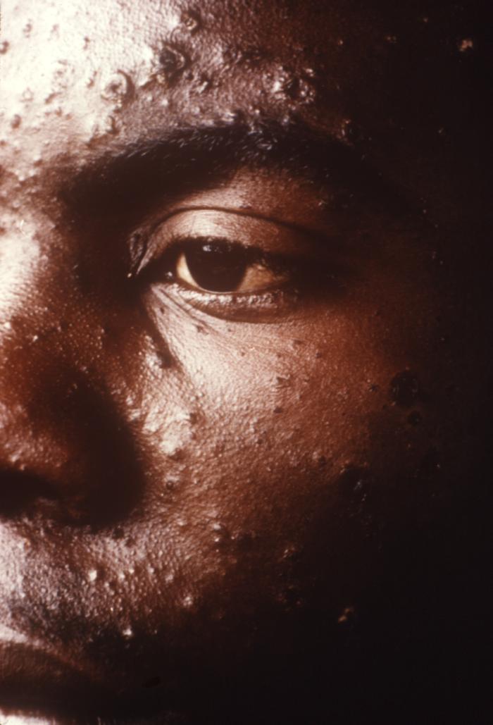
![Pathologic changes seen on the surface of the right unilateral side of this elderly male patient’s tongue and chin, represent a herpes outbreak due to the Varicella-zoster virus (VZV) pathogen. From Public Health Image Library (PHIL). [2]](https://www.wikidoc.org/images/b/b8/Chickenpox18.jpeg) Pathologic changes seen on the surface of the right unilateral side of this elderly male patient’s tongue and chin, represent a herpes outbreak due to the Varicella-zoster virus (VZV) pathogen. From Public Health Image Library (PHIL). [2]
Pathologic changes seen on the surface of the right unilateral side of this elderly male patient’s tongue and chin, represent a herpes outbreak due to the Varicella-zoster virus (VZV) pathogen. From Public Health Image Library (PHIL). [2] -
![Viewed from above, this image depicts a smallpox scab (left), and chickenpox scab (right) as a demonstration in comparative morphology. From Public Health Image Library (PHIL). [2]](https://www.wikidoc.org/images/a/ae/Chickenpox17.jpeg) Viewed from above, this image depicts a smallpox scab (left), and chickenpox scab (right) as a demonstration in comparative morphology. From Public Health Image Library (PHIL). [2]
Viewed from above, this image depicts a smallpox scab (left), and chickenpox scab (right) as a demonstration in comparative morphology. From Public Health Image Library (PHIL). [2] -
![Close-up of a maculopapular rash that was diagnosed as a crop of chickenpox lesions. From Public Health Image Library (PHIL). [2]](https://www.wikidoc.org/images/a/a8/Chickenpox16.jpeg) Close-up of a maculopapular rash that was diagnosed as a crop of chickenpox lesions. From Public Health Image Library (PHIL). [2]
Close-up of a maculopapular rash that was diagnosed as a crop of chickenpox lesions. From Public Health Image Library (PHIL). [2] -
![Lateral view of a 4 month-old infant’s face with a single varicella-zoster, otherwise known as chickenpox. From Public Health Image Library (PHIL). [2]](https://www.wikidoc.org/images/4/45/Chickenpox15.png) Lateral view of a 4 month-old infant’s face with a single varicella-zoster, otherwise known as chickenpox. From Public Health Image Library (PHIL). [2]
Lateral view of a 4 month-old infant’s face with a single varicella-zoster, otherwise known as chickenpox. From Public Health Image Library (PHIL). [2] -
![This anteroposterior (AP) radiograph revealed bilateral pulmonary infiltrates throughout the entirety of each lung field in the case of a child with leukemia, as well as chickenpox pneumonia. From Public Health Image Library (PHIL). [2]](https://www.wikidoc.org/images/0/0f/Chickenpox06.jpeg) This anteroposterior (AP) radiograph revealed bilateral pulmonary infiltrates throughout the entirety of each lung field in the case of a child with leukemia, as well as chickenpox pneumonia. From Public Health Image Library (PHIL). [2]
This anteroposterior (AP) radiograph revealed bilateral pulmonary infiltrates throughout the entirety of each lung field in the case of a child with leukemia, as well as chickenpox pneumonia. From Public Health Image Library (PHIL). [2] -
![Image depicts three mounted chickenpox scabs seen from the side revealing the superficiality of these scabs when morphologically compared to a smallpox scab. From Public Health Image Library (PHIL). [2]](https://www.wikidoc.org/images/0/09/Chickenpox04.jpeg) Image depicts three mounted chickenpox scabs seen from the side revealing the superficiality of these scabs when morphologically compared to a smallpox scab. From Public Health Image Library (PHIL). [2]
Image depicts three mounted chickenpox scabs seen from the side revealing the superficiality of these scabs when morphologically compared to a smallpox scab. From Public Health Image Library (PHIL). [2] -
![Volar surface of a patient’s left forearm, including the palmar surface of the left hand upon which you’ll note classic maculopapular rash of chickenpox. From Public Health Image Library (PHIL). [2]](https://www.wikidoc.org/images/0/09/Chickenpox03.jpeg) Volar surface of a patient’s left forearm, including the palmar surface of the left hand upon which you’ll note classic maculopapular rash of chickenpox. From Public Health Image Library (PHIL). [2]
Volar surface of a patient’s left forearm, including the palmar surface of the left hand upon which you’ll note classic maculopapular rash of chickenpox. From Public Health Image Library (PHIL). [2] -
![Right lateral surface of a patient’s right lower leg and foot with classic maculopapular rash of chickenpox. From Public Health Image Library (PHIL). [2]](https://www.wikidoc.org/images/5/50/Chickenpox02.jpeg) Right lateral surface of a patient’s right lower leg and foot with classic maculopapular rash of chickenpox. From Public Health Image Library (PHIL). [2]
Right lateral surface of a patient’s right lower leg and foot with classic maculopapular rash of chickenpox. From Public Health Image Library (PHIL). [2] -
![Right lateral surface of a patient’s right lower leg and foot with classic maculopapular rash of chickenpox. From Public Health Image Library (PHIL). [2]](https://www.wikidoc.org/images/7/72/VZV03.jpeg) Right lateral surface of a patient’s right lower leg and foot with classic maculopapular rash of chickenpox. From Public Health Image Library (PHIL). [2]
Right lateral surface of a patient’s right lower leg and foot with classic maculopapular rash of chickenpox. From Public Health Image Library (PHIL). [2] -
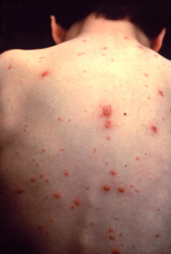
![Back of boy with chickenpox. From Public Health Image Library (PHIL). [2]](https://www.wikidoc.org/images/f/fd/VZV01.jpeg) Back of boy with chickenpox. From Public Health Image Library (PHIL). [2]
Back of boy with chickenpox. From Public Health Image Library (PHIL). [2] -
![Varicella From Public Health Image Library (PHIL). [2]](https://www.wikidoc.org/images/c/c8/Varicella_01.jpeg) Varicella From Public Health Image Library (PHIL). [2]
Varicella From Public Health Image Library (PHIL). [2] -
![Varicella From Public Health Image Library (PHIL). [2]](https://www.wikidoc.org/images/e/e7/Varicella_02.jpeg) Varicella From Public Health Image Library (PHIL). [2]
Varicella From Public Health Image Library (PHIL). [2] -
![Varicella From Public Health Image Library (PHIL). [2]](https://www.wikidoc.org/images/1/1a/Varicella_03.jpeg) Varicella From Public Health Image Library (PHIL). [2]
Varicella From Public Health Image Library (PHIL). [2] -
![Varicella From Public Health Image Library (PHIL). [2]](https://www.wikidoc.org/images/6/6e/Varicella_04.jpeg) Varicella From Public Health Image Library (PHIL). [2]
Varicella From Public Health Image Library (PHIL). [2] -
![Varicella From Public Health Image Library (PHIL). [2]](https://www.wikidoc.org/images/d/d4/Varicella_05.jpeg) Varicella From Public Health Image Library (PHIL). [2]
Varicella From Public Health Image Library (PHIL). [2] -
![Varicella From Public Health Image Library (PHIL). [2]](https://www.wikidoc.org/images/4/4d/Varicella_06.jpeg) Varicella From Public Health Image Library (PHIL). [2]
Varicella From Public Health Image Library (PHIL). [2] -
![Varicella From Public Health Image Library (PHIL). [2]](https://www.wikidoc.org/images/2/26/Varicella_07.jpeg) Varicella From Public Health Image Library (PHIL). [2]
Varicella From Public Health Image Library (PHIL). [2] -
![Varicella From Public Health Image Library (PHIL). [2]](https://www.wikidoc.org/images/6/61/Varicella_08.jpeg) Varicella From Public Health Image Library (PHIL). [2]
Varicella From Public Health Image Library (PHIL). [2] -
![Vasculitis leukocytoclasia. From Public Health Image Library (PHIL). [2]](https://www.wikidoc.org/images/4/47/Varicella_10.jpeg) Vasculitis leukocytoclasia. From Public Health Image Library (PHIL). [2]
Vasculitis leukocytoclasia. From Public Health Image Library (PHIL). [2] -
![Vasculitis leukocytoclasia. From Public Health Image Library (PHIL). [2]](https://www.wikidoc.org/images/8/8c/Varicella_11.jpeg) Vasculitis leukocytoclasia. From Public Health Image Library (PHIL). [2]
Vasculitis leukocytoclasia. From Public Health Image Library (PHIL). [2] -
![Vasculitis leukocytoclasia. From Public Health Image Library (PHIL). [2]](https://www.wikidoc.org/images/4/46/Varicella_12.jpeg) Vasculitis leukocytoclasia. From Public Health Image Library (PHIL). [2]
Vasculitis leukocytoclasia. From Public Health Image Library (PHIL). [2] -
![Vasculitis leukocytoclasia. From Public Health Image Library (PHIL). [2]](https://www.wikidoc.org/images/6/69/Varicella_13.jpeg) Vasculitis leukocytoclasia. From Public Health Image Library (PHIL). [2]
Vasculitis leukocytoclasia. From Public Health Image Library (PHIL). [2] -
![Vasculitis leukocytoclasia. From Public Health Image Library (PHIL). [2]](https://www.wikidoc.org/images/6/60/Varicella_14.jpeg) Vasculitis leukocytoclasia. From Public Health Image Library (PHIL). [2]
Vasculitis leukocytoclasia. From Public Health Image Library (PHIL). [2]
![Chickenpox lesions on the skin of this patient's left breast and arm on day 6 of the illness. From Public Health Image Library (PHIL). [2]](https://www.wikidoc.org/index.php/File%3AChickenpox36.jpeg)
![Chickenpox lesions on the skin of this patient's back and buttocks at day 6 of the illness. From Public Health Image Library (PHIL). [2]](https://www.wikidoc.org/index.php/File%3AChickenpox35.jpeg)
![Chickenpox lesions on the skin of this patient's breasts, arms, and torso at day 6 of the illness. From Public Health Image Library (PHIL). [2]](https://www.wikidoc.org/index.php/File%3AChickenpox34.jpeg)
![Patient with cervical skin lesions caused by chickenpox. From Public Health Image Library (PHIL). [2]](https://www.wikidoc.org/index.php/File%3AChickenpox33.jpeg)
![4-month old infant with skin lesions on his brow ridge due to chickenpox. From Public Health Image Library (PHIL). [2]](https://www.wikidoc.org/index.php/File%3AChickenpox32.jpeg)
![Patient had presented with chickenpox demonstrating the typical rash on day eight. From Public Health Image Library (PHIL). [2]](https://www.wikidoc.org/index.php/File%3AChickenpox29.jpeg)
![Patient developed palatal mucosal lesions due to chickenpox. From Public Health Image Library (PHIL). [2]](https://www.wikidoc.org/index.php/File%3AChickenpox28.jpeg)
![Vaccine recipient developed a secondary herpes infection adjacent to the vaccination site. From Public Health Image Library (PHIL). [2]](https://www.wikidoc.org/index.php/File%3AChickenpox27.jpeg)
![Pustulovesicular rash represents a generalized herpes outbreak due to the Varicella-zoster virus (VZV) pathogen. From Public Health Image Library (PHIL). [2]](https://www.wikidoc.org/index.php/File%3AChickenpox26.jpeg)
![Case of chickenpox. From Public Health Image Library (PHIL). [2]](https://www.wikidoc.org/index.php/File%3AChickenpox24.jpeg)
![Case of chickenpox. From Public Health Image Library (PHIL). [2]](https://www.wikidoc.org/index.php/File%3AChickenpox23.jpeg)
![Case of chickenpox. From Public Health Image Library (PHIL). [2]](https://www.wikidoc.org/index.php/File%3AChickenpox22.jpeg)
![Chickenpox lesions on a patient’s back, which were displaying the characteristic “cropping” distribution, or manifesting themselves in clusters. From Public Health Image Library (PHIL). [2]](https://www.wikidoc.org/index.php/File%3AChickenpox21.jpeg)
![Posterior view of a hospitalized man's neck, back and shoulders, who’d been assigned a bed in a smallpox ward, due to an initially misdiagnosed illness, which turned out to be chickenpox. From Public Health Image Library (PHIL). [2]](https://www.wikidoc.org/index.php/File%3AChickenpox20.jpeg)
![View of a patient’s thighs and upper legs, who’d been diagnosed with chickenpox. From Public Health Image Library (PHIL). [2]](https://www.wikidoc.org/index.php/File%3AChickenpox19.jpeg)
![Pathologic changes seen on the surface of the right unilateral side of this elderly male patient’s tongue and chin, represent a herpes outbreak due to the Varicella-zoster virus (VZV) pathogen. From Public Health Image Library (PHIL). [2]](https://www.wikidoc.org/index.php/File%3AChickenpox18.jpeg)
![Viewed from above, this image depicts a smallpox scab (left), and chickenpox scab (right) as a demonstration in comparative morphology. From Public Health Image Library (PHIL). [2]](https://www.wikidoc.org/index.php/File%3AChickenpox17.jpeg)
![Close-up of a maculopapular rash that was diagnosed as a crop of chickenpox lesions. From Public Health Image Library (PHIL). [2]](https://www.wikidoc.org/index.php/File%3AChickenpox16.jpeg)
![This anteroposterior (AP) radiograph revealed bilateral pulmonary infiltrates throughout the entirety of each lung field in the case of a child with leukemia, as well as chickenpox pneumonia. From Public Health Image Library (PHIL). [2]](https://www.wikidoc.org/index.php/File%3AChickenpox06.jpeg)
![Image depicts three mounted chickenpox scabs seen from the side revealing the superficiality of these scabs when morphologically compared to a smallpox scab. From Public Health Image Library (PHIL). [2]](https://www.wikidoc.org/index.php/File%3AChickenpox04.jpeg)
![Volar surface of a patient’s left forearm, including the palmar surface of the left hand upon which you’ll note classic maculopapular rash of chickenpox. From Public Health Image Library (PHIL). [2]](https://www.wikidoc.org/index.php/File%3AChickenpox03.jpeg)
![Right lateral surface of a patient’s right lower leg and foot with classic maculopapular rash of chickenpox. From Public Health Image Library (PHIL). [2]](https://www.wikidoc.org/index.php/File%3AChickenpox02.jpeg)
![Right lateral surface of a patient’s right lower leg and foot with classic maculopapular rash of chickenpox. From Public Health Image Library (PHIL). [2]](https://www.wikidoc.org/index.php/File%3AVZV03.jpeg)
![Back of boy with chickenpox. From Public Health Image Library (PHIL). [2]](https://www.wikidoc.org/index.php/File%3AVZV01.jpeg)
![Varicella From Public Health Image Library (PHIL). [2]](https://www.wikidoc.org/index.php/File%3AVaricella_01.jpeg)
![Varicella From Public Health Image Library (PHIL). [2]](https://www.wikidoc.org/index.php/File%3AVaricella_02.jpeg)
![Varicella From Public Health Image Library (PHIL). [2]](https://www.wikidoc.org/index.php/File%3AVaricella_03.jpeg)
![Varicella From Public Health Image Library (PHIL). [2]](https://www.wikidoc.org/index.php/File%3AVaricella_04.jpeg)
![Varicella From Public Health Image Library (PHIL). [2]](https://www.wikidoc.org/index.php/File%3AVaricella_05.jpeg)
![Varicella From Public Health Image Library (PHIL). [2]](https://www.wikidoc.org/index.php/File%3AVaricella_06.jpeg)
![Varicella From Public Health Image Library (PHIL). [2]](https://www.wikidoc.org/index.php/File%3AVaricella_07.jpeg)
![Varicella From Public Health Image Library (PHIL). [2]](https://www.wikidoc.org/index.php/File%3AVaricella_08.jpeg)
![Vasculitis leukocytoclasia. From Public Health Image Library (PHIL). [2]](https://www.wikidoc.org/index.php/File%3AVaricella_10.jpeg)
![Vasculitis leukocytoclasia. From Public Health Image Library (PHIL). [2]](https://www.wikidoc.org/index.php/File%3AVaricella_11.jpeg)
![Vasculitis leukocytoclasia. From Public Health Image Library (PHIL). [2]](https://www.wikidoc.org/index.php/File%3AVaricella_12.jpeg)
![Vasculitis leukocytoclasia. From Public Health Image Library (PHIL). [2]](https://www.wikidoc.org/index.php/File%3AVaricella_13.jpeg)
![Vasculitis leukocytoclasia. From Public Health Image Library (PHIL). [2]](https://www.wikidoc.org/index.php/File%3AVaricella_14.jpeg)
Unvaccinated Individuals
-
 Girl with a secondary skin infection due to chickenpox.
 -
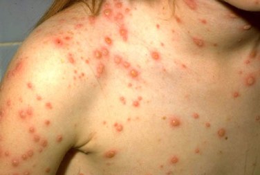
 Chickenpox in an unvaccinated child.
 -
 Chickenpox in an unvaccinated adult.
 -
 Chickenpox in unvaccinated adult.
 -
 Chickenpox in unvaccinated child.


Vaccinated Individuals
-
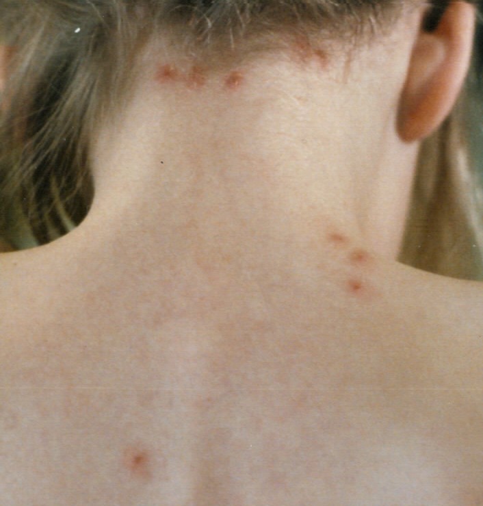
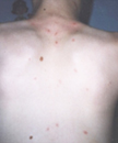
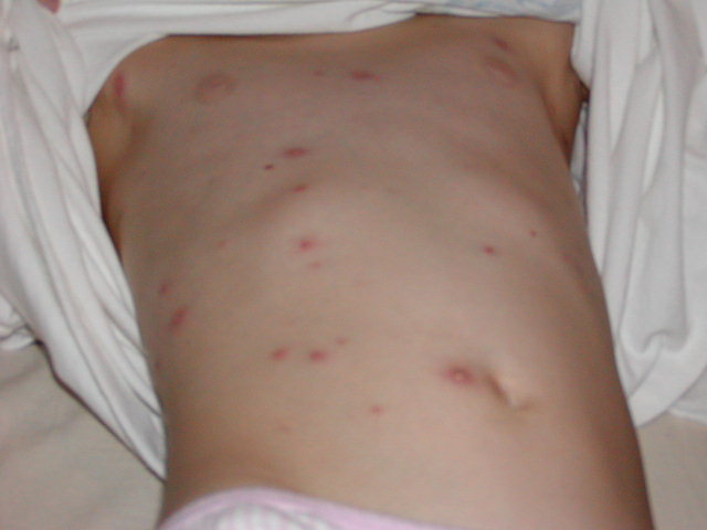 Image of Breakthrough Chickenpox: Back of child with breakthrough varicella.
 -
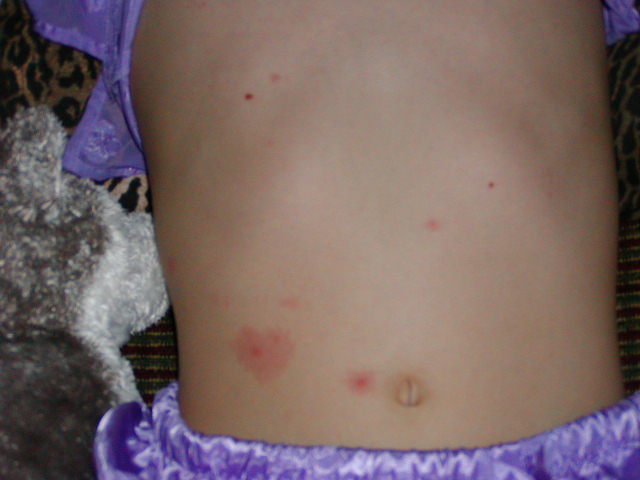 Image of Breakthrough Chickenpox: Back of child with breakthrough varicella.
 -
 The skin lesions of breakthrough varicella can be macular rather than vesicular. They are rarely bullous or hemorrhagic, and residual scarring is less common.
 -
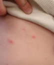 Breakthrough varicella on the abdomen of a vaccinated child.
 -
 Breakthrough varicella on the back of a vaccinated child.


References
References
- ↑ 1.0 1.1 Schraufnagel DE, Becker RP, Balaan M, Schmid A, Claypool W (1989). “Silver staining of Pneumocystis carinii in the rat’s lung”. J Infect. 18 (1): 39–44. PMID 2464648.
- ↑ 2.00 2.01 2.02 2.03 2.04 2.05 2.06 2.07 2.08 2.09 2.10 2.11 2.12 2.13 2.14 2.15 2.16 2.17 2.18 2.19 2.20 2.21 2.22 2.23 2.24 2.25 2.26 2.27 2.28 2.29 2.30 2.31 2.32 2.33 2.34 2.35 2.36 2.37 “Public Health Image Library (PHIL)”.
Looking for the patient version?
© 2026 MyEClinic – IFTM Institut für Telematik in der Medizin GmbH