
Choroid
Editor-In-Chief: C. Michael Gibson, M.S., M.D. [1]
The choroid, also known as the choroidea or choroid coat, is the vascular layer of the eye lying between the retina and the sclera, with a thickness about 0.5 mm. The choroid provides oxygen and nourishment to the outer layers of the retina [2].
Along with the ciliary body and iris, the choroid forms the uveal tract. In humans and other primates, darkly colored melanin pigment in the choroid helps limit reflections within the eye that would potentially result in the perception of confusing images. Poor vision frequently results from lack of this pigmentation in human albinos. By contrast, the choroid of many other animals contains reflective materials that help to collect light in dim situations; this is one type of tapetum lucidum.
The red eye effect on photos is caused by the reflection of light from choroid. It appears red because of the choroid’s blood vessels.
Layers
Layers
The structure of the choroid is generally divided into four layers:
- Haller’s layer – outermost layer of the choroid consisting of larger diameter blood vessels
- Sattler’s layer – layer of medium diameter blood vessels
- Choriocapillaris – layer of capillaries
- Bruch’s membrane – innermost layer of the choroid
Additional images
Additional images
-
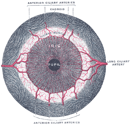 Iris, front view.
 -
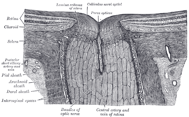 The terminal portion of the optic nerve and its entrance into the eyeball, in horizontal section.
 -
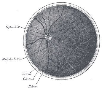 The interior of the posterior half of the left eyeball.


External links
External links
- Histology image: 08008loa – Histology Learning System at Boston University
ca:Coroide de:Chorioidea el:Χοριοειδής χιτώνας he:שכבה דמית lt:Gyslainė nl:Vaatvlies sk:Cievovka
Looking for the patient version?
© 2026 MyEClinic – IFTM Institut für Telematik in der Medizin GmbH