
Bone fracture

For patient information click here
Editor-In-Chief: C. Michael Gibson, M.S., M.D. [1]; Associate Editor(s)-in-Chief: Mohammadmain Rezazadehsaatlou[2].
Synonyms and keywords: Fracture of bone
Overview
Editor-In-Chief: C. Michael Gibson, M.S., M.D. [1]
Overview
A bone fracture is a medical condition in which a bone is cracked or broken. While many fractures are the result of high force impact or stress, bone fracture can also occur as a result of certain medical conditions that weaken the bones, such as osteoporosis, certain types of cancer or osteogenesis imperfecta.
Historical Perspective
Any type of bone break is a fracture. The word break is not used in formal orthopedic terminology.
Risk Factors
Fractures commonly occur because of falls or sports injuries. Additionally, overuse can cause stress fractures, which are very small cracks in the bone. Therefore, those who participate in rigorous physical activities or in high risk activities are more likely to get a fracture. Another common cause of fractures is osteoporosis, which causes weakening of the bones. People with this condition, especially the elderly, are at higher risk of fractures due to falls since their bones cannot withstand as much stress.
Diagnosis
A bone fracture can be diagnosed clinically based on the medical history given and the physical examination performed. Imaging by X-ray is often performed to view the bone suspected of being fractured. In situations where x-ray alone is insufficient, a computed tomograph (CT scan) may be performed.
X Ray
At the hospital, closed fractures are diagnosed by taking an X-ray photograph of the injury.
References
Historical Perspective
For patient information click here
Editor-In-Chief: C. Michael Gibson, M.S., M.D. [1]; Associate Editor(s)-in-Chief: Mohammadmain Rezazadehsaatlou[2].
Overview
Any type of bone break is a fracture. The word break is not used in formal orthopedic terminology.
Bone fracture historical perspective
3000 BC: The earliest examples of fractures and related splinted before death found at Naga-ed-der (about 100 miles north of Luxor in Egypt.
Until 19th century: Splintage with wooden apparatus was the only available therapeutic option.
Then the new techniques became available.
References
Classification
Editor-In-Chief: C. Michael Gibson, M.S., M.D. [1]
Classification
In orthopedic medicine, fractures are classified as closed or open (compound) and simple or multi-fragmentary (formerly comminuted).
- Closed fractures are those in which the skin is intact, while open (compound) fractures involve wounds that communicate with the fracture and may expose bone to contamination. Open injuries carry an elevated risk of infection; they require antibiotic treatment and usually urgent surgical treatment (debridement). This involves removal of all dirt, contamination, and dead tissue.
- Simple fractures are fractures that only occur along one line, splitting the bone into two pieces, while multi-fragmentary fractures involve the bone splitting into multiple pieces. A simple, closed fracture is much easier to treat and has a much better prognosis than an open, contaminated fracture. Other considerations in fracture care are displacement (fracture gap) and angulation. If angulation or displacement is large, reduction (manipulation) of the bone may be required and, in adults, frequently requires surgical care. These injuries may take longer to heal than injuries without displacement or angulation.
Another type of bone fracture is a compression fracture. An example of a compression fracture is when the front portion of a vertebra in the spine collapses due to osteoporosis, a medical condition which causes bones to become brittle and susceptible to fracture (with or without trauma).
Other types of fracture are:
- Complete Fracture- A fracture in which bone fragments separate completely.
- Incomplete Fracture- A fracture in which the bone fragments are still partially joined.
- Linear Fracture- A fracture that is parallel to the bone’s long axis.
- Transverse Fracture- A fracture that is at a right angle to the bone’s long axis.
- Oblique Fracture- A fracture that is diagonal to a bone’s long axis.
- Compression Fracture-A fracture that usually occurs in the vertebrae.
- Spiral Fracture- A fracture where at least one part of the bone has been twisted.
- Comminuted Fracture- A fracture causing many fragments.
- Compacted Fracture- A fracture caused when bone fragments are driven into each other
- Open Fracture- A fracture when the bone reaches the skin
OTA Classification
The Orthopaedic Trauma Association, an association for Orthopaedic surgeons, devised an elaborate classification system to describe the injury accurately and guide treatment.[1][2] There are five parts to the code:
- Bone: Description of a fracture starts by naming the bone
- Location: the part of the bone involved (e.g. shaft of the femur).
- 1) Proximal
- 2) Diaphyseal
- 3) Distal
- Type: It is important to note whether the fracture is simple or multifragmentary and whether it is closed or open.
- A = Simple fracture
- B = Wedge fracture
- C = Complex fracture
- Group: The geometry of the fracture is also described by terms such as transverse, oblique, spiral, or segmental.
- Subgroup: Other features of the fracture are described in terms of displacement, angulation and shortening. A stable fracture is one which is likely to stay in a good (functional) position while it heals; an unstable one is likely to shorten, angulate or rotate before healing and lead to poor function in the long term.
Other Classification Systems
There are other systems used to classify different types of bone fractures:
References
- ↑ “Fracture and dislocation compendium. Orthopaedic Trauma Association Committee for Coding and Classification” (pdf). J Orthop Trauma. 10 Suppl 1: v–ix, 1–154. 1996. PMID 8814583. Retrieved 2007-11-28.
- ↑ “Orthopaedic Trauma Association/ Committee for Coding and Classification: Fracture and Dislocation Compendium”. Orthopaedic Trauma Association. Retrieved 2007-11-28.
- ↑ Mourad L (1997). “Neer classification of fractures of the proximal humerus”. Orthop Nurs. 16 (2): 76. PMID 9155417.
- ↑ “eMedicine – Proximal Humerus Fractures : Article by Mark Frankle, MD”. Retrieved 2007-12-15.
- ↑ Template:GPnotebook
- ↑ “Seinsheimer’s Classification of Subtrochanteric Frxs – Wheeless’ Textbook of Orthopaedics”. Retrieved 2007-12-15.
Pathophysiology
Please help WikiDoc by adding content here. It’s easy! Click here to learn about editing.
References
Causes
Editor-In-Chief: C. Michael Gibson, M.S., M.D. [1]
Causes
The following are common causes of broken bones:
- Fall from a height
- Motor vehicle accidents
- Direct blow
- Child abuse
- Drugs– ibandronate, Isotretinoin, Pantoprazole, Rabeprazole, Saxagliptin
- Repetitive forces, such as those caused by running, can cause stress fractures of the foot, ankle, tibia, or hip
References
Differentiating Bone fracture from other Diseases
Please help WikiDoc by adding content here. It’s easy! Click here to learn about editing.
References
Epidemiology and Demographics
Please help WikiDoc by adding content here. It’s easy! Click here to learn about editing.
References
Risk Factors
Editor-In-Chief: C. Michael Gibson, M.S., M.D. [1]
Please help WikiDoc by adding content here. It’s easy! Click here to learn about editing.
Overview
Fractures commonly occur because of falls or sports injuries. Additionally, overuse can cause stress fractures, which are very small cracks in the bone. Therefore, those who participate in rigorous physical activities or in high risk activities are more likely to get a fracture. Another common cause of fractures is osteoporosis, which causes weakening of the bones. People with this condition, especially the elderly, are at higher risk of fractures due to falls since their bones cannot withstand as much stress.
References
Natural History, Complications and Prognosis
Editor-In-Chief: C. Michael Gibson, M.S., M.D. [1]
Natural History
Bone Healing
The natural process of healing a fracture starts when the injured bone and surrounding tissues bleed. The blood coagulates to form a blood clot situated between the broken fragments. Within a few days blood vessels grow into the jelly-like matrix of the blood clot. The new blood vessels bring white blood cells to the area, which gradually remove the non-viable material. The blood vessels also bring fibroblasts in the walls of the vessels and these multiply and produce collagen fibers. In this way the blood clot is replaced by a matrix of collagen. Collagen’s rubbery consistency allows bone fragments to move only a small amount unless severe or persistent force is applied.
At this stage, some of the fibroblasts begin to lay down bone matrix (calcium hydroxyapatite) in the form of insoluble crystals. This mineralization of the collagen matrix stiffens it and transforms it into bone. In fact, bone is a mineralized collagen matrix; if the mineral is dissolved out of bone, it becomes rubbery. Healing bone callus is on average sufficiently mineralized to show up on X-ray within 6 weeks in adults and less in children. This initial “woven” bone does not have the strong mechanical properties of mature bone. By a process of remodeling, the woven bone is replaced by mature “lamellar” bone. The whole process can take up to 18 months, but in adults the strength of the healing bone is usually 80% of normal by 3 months after the injury.
Several factors can help or hinder the bone healing process. For example, any form of nicotine hinders the process of bone healing, and adequate nutrition (including calcium intake) will help the bone healing process. Weight-bearing stress on bone, after the bone has healed sufficiently to bear the weight, also builds bone strength. Although there are theoretical concerns about NSAIDs slowing the rate of healing, there is not enough evidence to warrant withholding the use of this type analgesic in simple fractures.
-
 X-ray showing a healed tibia fracture with pins.
 -
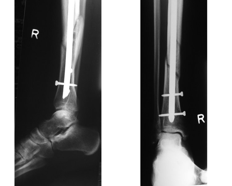 X-ray showing fractured tibia and pinning.


Complications
Some fractures can lead to serious complications including a condition known as compartment syndrome. If not treated, compartment syndrome can result in amputation of the affected limb. Other complications may include non-union, where the fractured bone fails to heal or mal-union, where the fractured bone heals in a deformed manner.
In children, whose bones are still developing, there are risks of either a growth plate injury or a greenstick fracture.
- A greenstick fracture occurs due to mechanical failure on the tension side. That is, since the bone is not as brittle as it would be in an adult, it does not completely fracture, but rather exhibits bowing without complete disruption of the bone’s cortex in the surface opposite the applied force.
- Growth plate injuries, as in Salter-Harris fractures, require careful treatment and accurate reduction to make sure that the bone continues to grow normally.
- Plastic deformation of the bone, in which the bone permanently bends but does not break, is also possible in children. These injuries may require an osteotomy (bone cut) to realign the bone if it is fixed and cannot be realigned by closed methods.
- Certain fractures are known to occur mainly in the pediatric age group, such as fracture of the clavicle and supracondylar fracture of the humerus.
Prognosis
For most fractures, the whole process can take up to 18 months, but in adults the strength of the healing bone is usually 80% of normal by 3 months after the injury.
References
Diagnosis
Diagnosis
History and Symptoms | Physical Examination | Laboratory Findings | X Ray | CT | MRI | Ultrasound | Other Imaging Findings | Other Diagnostic Studies
Treatment
Treatment
Medical Therapy | Surgery | Primary Prevention | Secondary Prevention | Cost-Effectiveness of Therapy | Future or Investigational Therapies
Related Chapters
Related Chapters
ar:كسر عظم ca:Fractura (medicina) de:Knochenbruch el:Κάταγμα eo:Frakturo (medicino) gl:Fractura ko:골절 it:Frattura (medicina) he:שבר (רפואה) lt:Kaulo lūžimas nl:Botbreuk no:Beinbrudd qu:Tullup’aki sk:Zlomenina kosti fi:Luunmurtuma sv:Fraktur (medicin) uk:Переломи кісток
Looking for the patient version?
© 2026 MyEClinic – IFTM Institut für Telematik in der Medizin GmbH