
De Quervain's thyroiditis other diagnostic studies
Editor-In-Chief: C. Michael Gibson, M.S., M.D. [1]; Associate Editor(s)-in-Chief: Furqan M M. M.B.B.S[2]
Overview
Overview
The histological analysis in de Quervain’s thyroiditis may show the destruction of the follicular epithelium, loss of the follicular integrity, and infiltration of inflammatory cells. Fine needle aspiration cytology helps to differentiate between the benign and malignant nodules.
Other Diagnostic Studies
Other Diagnostic Studies
Microscopic Pathology
Microscopic findings suggesting de Quervain’s thyroiditis are as followings:[1]
- Granuloma comprises of colloid, small lymphocytes, neutrophils, and macrophages with or without epithelioid features
- Destruction of the follicular epithelium
- Loss of the follicular integrity
- Patchy distribution of non-caseous granulomas
Fine needle aspiration cytology
Fine needle aspiration is usually done under ultrasound guidance and the sample is sent for cytology. It helps to differentiate benign thyroid nodules from the malignant lesions.[2][3]
Gallery
Gallery
-
 Histology of De Quervain’s thyroiditis; Granuloma (By Nephron – Own work, CC BY-SA 3.0, https://commons.wikimedia.org/w/index.php?curid=18491382)
 -
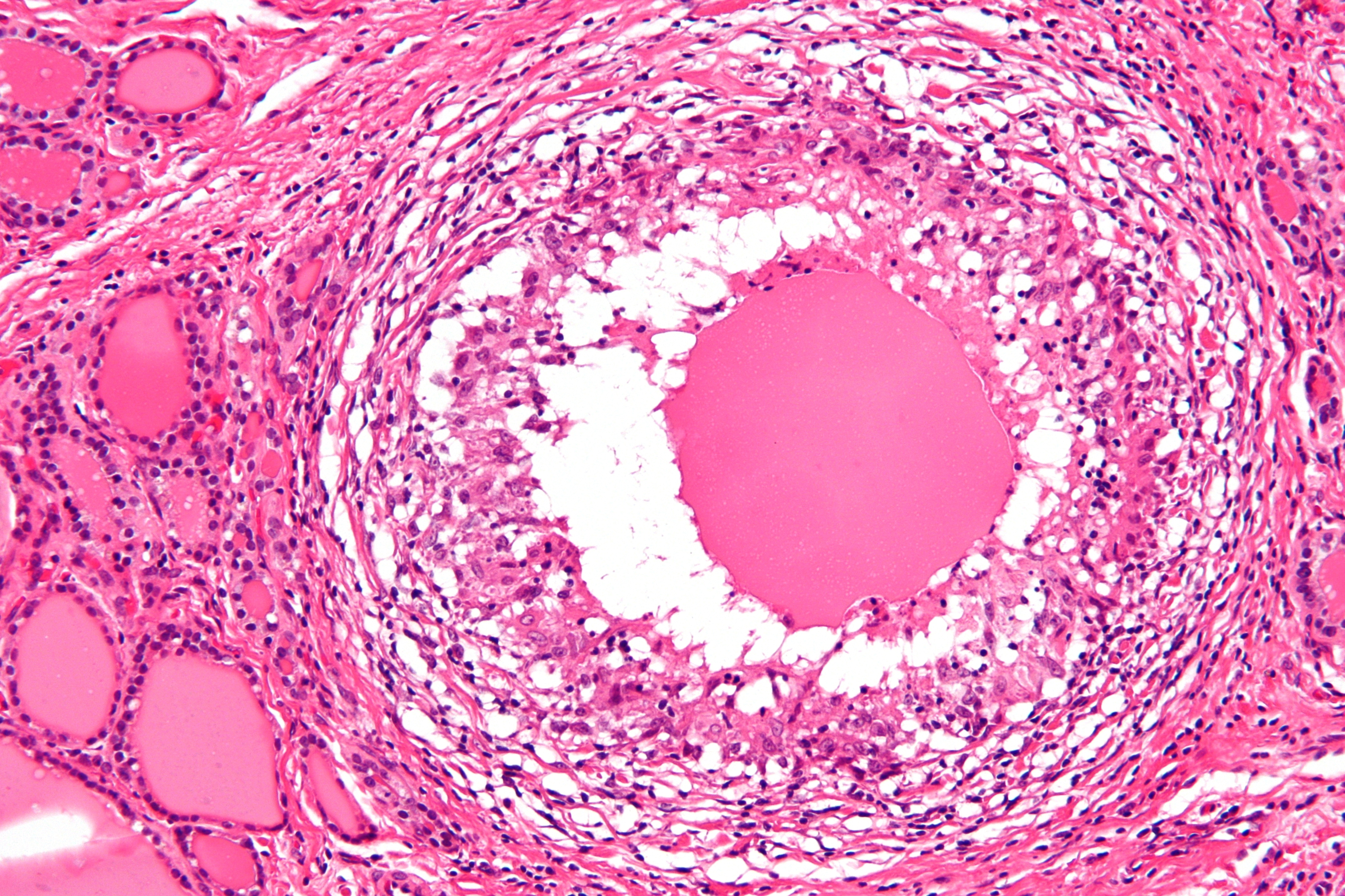 Histology of De Quervain’s thyroiditis; Granuloma (By Nephron – Own work, CC BY-SA 3.0, https://commons.wikimedia.org/w/index.php?curid=18491421)
 -
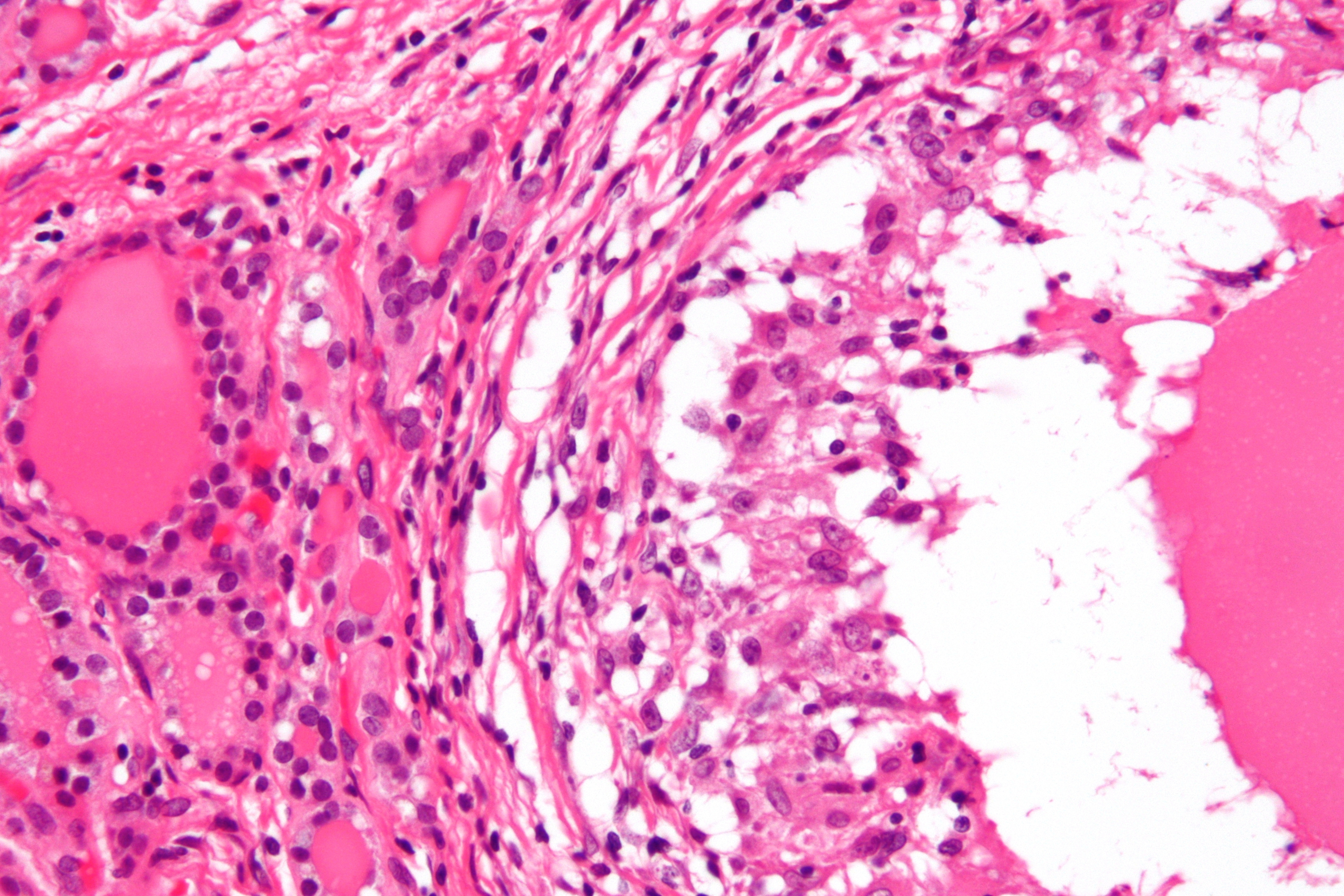 Histology of De Quervain’s thyroiditis; Granuloma (By Nephron – Own work, CC BY-SA 3.0, https://commons.wikimedia.org/w/index.php?curid=18491421)
 -
![FNA smear of De Quervain's thyroiditis; Inflammatory rich smear with giant cells and probable granulomata. (Case courtesy of Dr Andrew Ryan, <a href="https://radiopaedia.org/">Radiopaedia.org</a>. From the case <a href="https://radiopaedia.org/cases/17052">rID: 17052</a>)]])](https://www.wikidoc.org/images/1/1c/Subacute-de-quervains-thyroiditis_%283%29.jpg) FNA smear of De Quervain’s thyroiditis; Inflammatory rich smear with giant cells and probable granulomata. (Case courtesy of Dr Andrew Ryan, <a href=”https://radiopaedia.org/“>Radiopaedia.org</a>. From the case <a href=”https://radiopaedia.org/cases/17052“>rID: 17052</a>)]])
FNA smear of De Quervain’s thyroiditis; Inflammatory rich smear with giant cells and probable granulomata. (Case courtesy of Dr Andrew Ryan, <a href=”https://radiopaedia.org/“>Radiopaedia.org</a>. From the case <a href=”https://radiopaedia.org/cases/17052“>rID: 17052</a>)]]) -
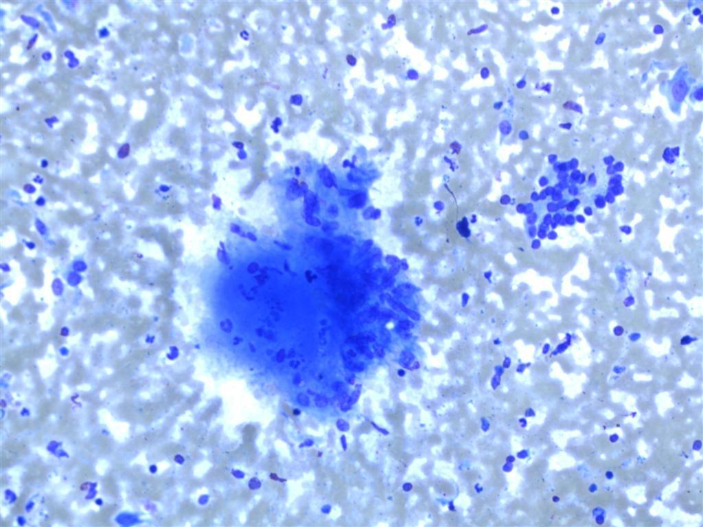 FNA smear of De Quervain’s thyroiditis; ; Cluster of epithelioid histiocytes with associated inflammatory cells and debris and group of benign follicular epithelial cells. (Case courtesy of Dr Andrew Ryan, <a href=”https://radiopaedia.org/“>Radiopaedia.org</a>. From the case <a href=”https://radiopaedia.org/cases/17052“>rID: 17052</a>)


![FNA smear of De Quervain's thyroiditis; Inflammatory rich smear with giant cells and probable granulomata. (Case courtesy of Dr Andrew Ryan, <a href="https://radiopaedia.org/">Radiopaedia.org</a>. From the case <a href="https://radiopaedia.org/cases/17052">rID: 17052</a>)]])](https://www.wikidoc.org/index.php/File%3ASubacute-de-quervains-thyroiditis_%283%29.jpg)

References
References
- ↑ Kojima M, Nakamura S, Oyama T, Sugihara S, Sakata N, Masawa N (2002). “Cellular composition of subacute thyroiditis. an immunohistochemical study of six cases”. Pathol. Res. Pract. 198 (12): 833–7. doi:10.1078/0344-0338-00344. PMID 12608662.
- ↑ “Thyroiditis — NEJM”.
- ↑ Fatourechi V, Aniszewski JP, Fatourechi GZ, Atkinson EJ, Jacobsen SJ (2003). “Clinical features and outcome of subacute thyroiditis in an incidence cohort: Olmsted County, Minnesota, study”. J. Clin. Endocrinol. Metab. 88 (5): 2100–5. doi:10.1210/jc.2002-021799. PMID 12727961.
Looking for the patient version?
© 2026 MyEClinic – IFTM Institut für Telematik in der Medizin GmbH