

Health Dictionary
Reliable, easy-to-read patient information on common medical conditions — written in plain language, free of jargon, and grounded in current evidence. Browse the most frequent diagnoses, understand what they mean for everyday life, and learn which steps and questions help you have a focused conversation with your doctor.
Epiglottic cartilage
Introduction
Overview
Overview
The epiglottis is a lid-like flap of elastic cartilage tissue covered with a mucus membrane, attached to the root of the tongue. It projects obliquely upwards behind the tongue and the hyoid bone.
Anatomy and function
Anatomy and function
The epiglottis guards the entrance of the glottis, the opening between the vocal folds.
It is normally pointed upward, but during swallowing, elevation of the hyoid bone draws the larynx upward; as a result, the epiglottis folds down to a more horizontal position. In this manner it prevents food from going into the trachea and instead directs it to the esophagus, which is more posterior.
The epiglottis is one of three large cartilaginous structures that make up the larynx (voice box).
Clinical significance
Clinical significance
Reflexes
The glossopharyngeal nerve (CN IX) sends fibers to the upper epiglottis that contribute to the afferent limb of the gag reflex. The superior laryngeal branch of the vagus nerve (CN X) sends fibers to the lower epiglottis that contribute to the afferent limb of the cough reflex. [1]
Infection of the epiglottis
In children, the epiglottis will occasionally become infected with Haemophilus influenzae or Streptococcus pneumoniae bacteria. Although easily treated, this condition is a medical emergency because without treatment the epiglottis may swell and block the trachea, causing massive inflammation. This condition has become rare in countries where vaccination against Haemophilus influenzae (HIB) is administered.
Additional images
Additional images
-
 Larynx
 -
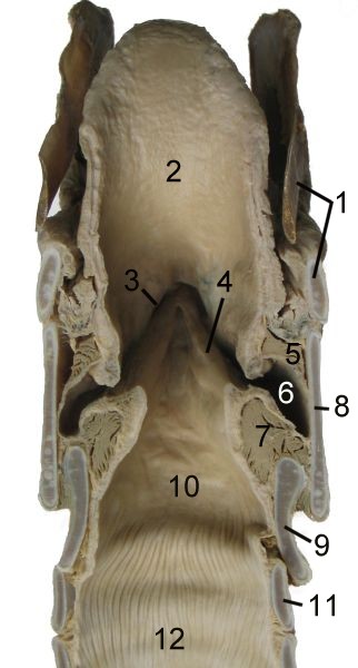 Cut through the larynx of a horse
 -
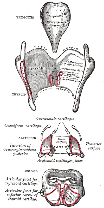 The cartilages of the larynx. Posterior view.
 -
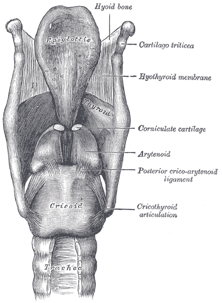 Ligaments of the larynx. Posterior view.
 -
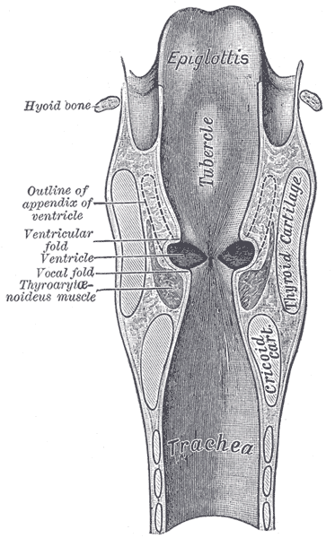 Coronal section of larynx and upper part of trachea.
 -
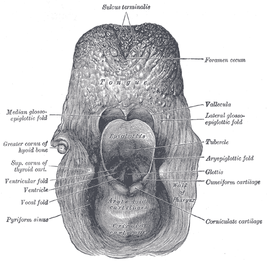 The entrance to the larynx, viewed from behind.
 -
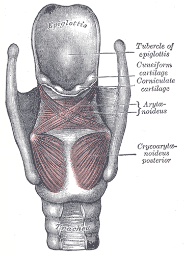 Muscles of larynx. Posterior view.
 -
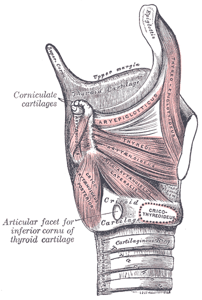 Muscles of larynx. Side view. Right lamina of thyroid cartilage removed.
 -
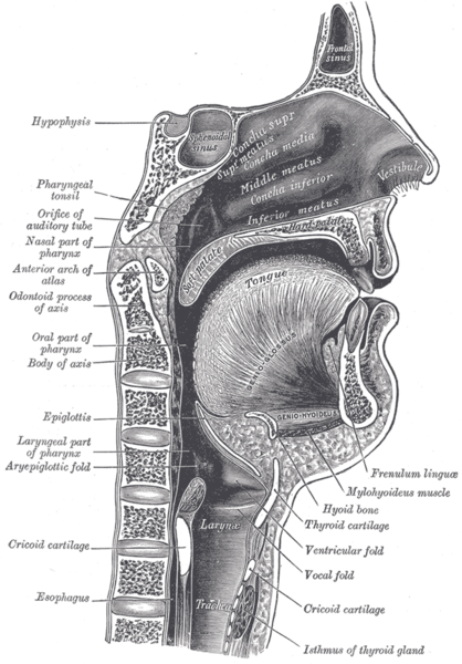 Sagittal section of nose mouth, pharynx, and larynx.


