
First pharyngeal arch
Editor-In-Chief: C. Michael Gibson, M.S., M.D. [1]
Overview
Overview
The first branchial arch, also called the first pharyngeal arch and mandibular arch, is the first of six branchial arches that develops in fetal life. It is located between the stomodeum and the first pharyngeal groove.
Processes
Processes
This arch divides into a maxillary process and a mandibular process, giving rise to structures including the bones of the lower two-thirds of the face and the jaw. The maxillary process becomes the maxilla (or upper jaw), and palate while the mandibular process becomes the lower jaw. This arch also gives rise to the muscles of mastication.
Innervation of the two processes of the first branchial arch is provided by two distinct branches of the trigeminal nerve (CN V),[1] the mandibular and maxillary branches. The artery of the first arch is the first aortic arch,[2] which partially persists as the maxillary artery.
Meckel’s cartilage
Meckel’s cartilage
Meckel’s cartilage forms in the mesoderm of the mandibular process and eventually regresses to form the incus and malleus of the middle ear; the anterior ligament of the malleus and the sphenomandibular ligament. The mandible or lower jaw forms by intramembranous ossification using Meckel’s cartilage as a ‘template’, but the mandible does not arise from direct ossification of Meckel’s cartilage.
Derivatives of the first arch
Derivatives of the first arch
Derivatives of the first arch:
- Ectodermal and endodermal
- mucous membrane and glands of the anterior two thirds of the tongue
- Mesodermal
- muscles of mastication (chewing)
- masseter
- pterygoid muscles
- temporalis muscles
- mylohyoid muscle
- digastric muscle, anterior belly
- tensor palati muscle
- tensor tympani muscle
- muscles of mastication (chewing)
Additional images
Additional images
-
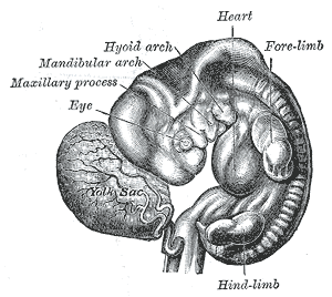 Human embryo from thirty-one to thirty-four days.
 -
 Head end of human embryo, about the end of the fourth week.
 -
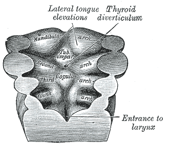 Floor of pharynx of embryo between 18 and 21 days.
 -
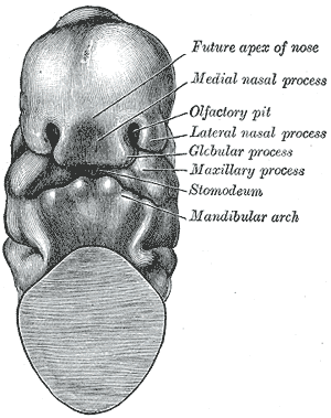 Head end of human embryo of about thirty to thirty-one days.
 -
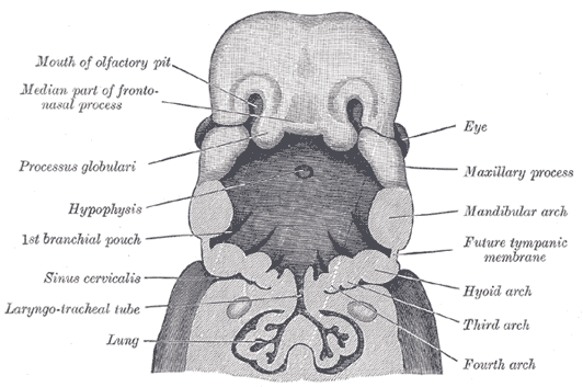 The head and neck of a human embryo thirty-two days old, seen from the ventral surface.


References
References
External links
External links
- Template:EmbryologyUNC
- Overview at University of Newcastle
- Overview at Howard University
Looking for the patient version?
© 2026 MyEClinic – IFTM Institut für Telematik in der Medizin GmbH