
List of distinct cell types in the adult human body
Editor-In-Chief: C. Michael Gibson, M.S., M.D. [1]
There are about 210 distinct human cell types.
Keratinizing epithelial cells
Keratinizing epithelial cells
- Epidermal keratinocyte (differentiating epidermal cell)
- Epidermal basal cell (stem cell)
- Keratinocyte of fingernails and toenails
- Nail bed basal cell (stem cell)
- Medullary hair shaft cell
- Cortical hair shaft cell
- Cuticular hair shaft cell
- Cuticular hair root sheath cell
- Hair root sheath cell of Huxley’s layer
- Hair root sheath cell of Henle’s layer
- External hair root sheath cell
- Hair matrix cell (stem cell)
Wet stratified barrier epithelial cells
Wet stratified barrier epithelial cells
- Surface epithelial cell of stratified squamous epithelium of cornea, tongue, oral cavity, esophagus, anal canal, distal urethra and vagina
- basal cell (stem cell) of epithelia of cornea, tongue, oral cavity, esophagus, anal canal, distal urethra and vagina
- Urinary epithelium cell (lining urinary bladder and urinary ducts)
Exocrine secretory epithelial cells
Exocrine secretory epithelial cells
- Salivary gland mucous cell (polysaccharide-rich secretion)
- Salivary gland serous cell (glycoprotein enzyme-rich secretion)
- Von Ebner’s gland cell in tongue (washes taste buds)
- Mammary gland cell (milk secretion)
- Lacrimal gland cell (tear secretion)
- Ceruminous gland cell in ear (wax secretion)
- Eccrine sweat gland dark cell (glycoprotein secretion)
- Eccrine sweat gland clear cell (small molecule secretion)
- Apocrine sweat gland cell (odoriferous secretion, sex-hormone sensitive)
- Gland of Moll cell in eyelid (specialized sweat gland)
- Sebaceous gland cell (lipid-rich sebum secretion)
- Bowman’s gland cell in nose (washes olfactory epithelium)
- Brunner’s gland cell in duodenum (enzymes and alkaline mucus)
- Seminal vesicle cell (secretes seminal fluid components, including fructose for swimming sperm)
- Prostate gland cell (secretes seminal fluid components)
- Bulbourethral gland cell (mucus secretion)
- Bartholin’s gland cell (vaginal lubricant secretion)
- Gland of Littre cell (mucus secretion)
- Uterus endometrium cell (carbohydrate secretion)
- Isolated goblet cell of respiratory and digestive tracts (mucus secretion)
- Stomach lining mucous cell (mucus secretion)
- Gastric gland zymogenic cell (pepsinogen secretion)
- Gastric gland oxyntic cell (hydrochloric acid secretion)
- Pancreatic acinar cell (bicarbonate and digestive enzyme secretion)
- Paneth cell of small intestine (lysozyme secretion)
- Type II pneumocyte of lung (surfactant secretion)
- Clara cell of lung
Hormone secreting cells
Hormone secreting cells
- Anterior pituitary cells
- Intermediate pituitary cell, secreting melanocyte-stimulating hormone
- Magnocellular neurosecretory cells
- secreting oxytocin
- secreting vasopressin
- Gut and respiratory tract cells secreting serotonin
- secreting endorphin
- secreting somatostatin
- secreting gastrin
- secreting secretin
- secreting cholecystokinin
- secreting insulin
- secreting glucagon
- secreting bombesin
- Thyroid gland cells
- Parathyroid gland cells
- Adrenal gland cells
- chromaffin cells
- secreting steroid hormones (mineralcorticoids and gluco corticoids)
- Leydig cell of testes secreting testosterone
- Theca interna cell of ovarian follicle secreting estrogen
- Corpus luteum cell of ruptured ovarian follicle secreting progesterone
- Juxtaglomerular cell (renin secretion)
- Macula densa cell of kidney
- Peripolar cell of kidney
- Mesangial cell of kidney
(Gut, Exocrine Glands and Urogenital Tract)
(Gut, Exocrine Glands and Urogenital Tract)
Metabolism and storage cells
Metabolism and storage cells
- Hepatocyte (liver cell)
- White fat cell
- Brown fat cell
- Liver lipocyte
Barrier function cells (Lung, Gut, Exocrine Glands and Urogenital Tract)
Barrier function cells (Lung, Gut, Exocrine Glands and Urogenital Tract)
- Type I pneumocyte (lining air space of lung)
- Pancreatic duct cell (centroacinar cell)
- Nonstriated duct cell (of sweat gland, salivary gland, mammary gland, etc.)
- Kidney glomerulus parietal cell
- Kidney glomerulus podocyte
- Loop of Henle thin segment cell (in kidney)
- Kidney collecting duct cell
- Duct cell (of seminal vesicle, prostate gland, etc.)


Template:WikiDoc Cardiology News
Editor-In-Chief: C. Michael Gibson, M.S., M.D. [1]
The lung is the essential respiration organ in air-breathing vertebrates, the most primitive being the lungfish. Its principal function is to transport oxygen from the atmosphere into the bloodstream, and to release carbon dioxide from the bloodstream into the atmosphere. This exchange of gases is accomplished in the mosaic of specialized cells that form millions of tiny, exceptionally thin-walled air sacs called alveoli. The lungs also have non respiratory functions.
Medical terms related to the lung often begin with pulmo-, from the Latin pulmonarius (“of the lungs”), or with pneumo- (from Greek πνεύμω “lung”)[2][3]
Respiratory function

Energy production from aerobic respiration requires oxygen and glucose and produces carbon dioxide as a waste product, creating a need for an efficient means of oxygen delivery to cells and excretion of carbon dioxide from cells. In small organisms, such as single-celled bacteria, this process of gas exchange can take place entirely by simple diffusion. In larger organisms, this is not possible; only a small proportion of cells are close enough to the surface for oxygen from the atmosphere to enter them through diffusion. Two major adaptations made it possible for organisms to attain great multicellularity: an efficient circulatory system that conveyed gases to and from the deepest tissues in the body, and a large, internalized respiratory system that centralized the task of obtaining oxygen from the atmosphere and bringing it into the body, whence it could rapidly be distributed to all the circulatory system.
In air-breathing vertebrates, respiration occurs in a series of steps. Air is brought into the animal via the airways — in reptiles, birds and mammals this often consists of the nose; the pharynx; the larynx; the trachea (also called the windpipe); the bronchi and bronchioles; and the terminal branches of the respiratory tree. The lungs of mammals are a rich lattice of alveoli, which provide an enormous surface area for gas exchange. A network of fine capillaries allows transport of blood over the surface of alveoli. Oxygen from the air inside the alveoli diffuses into the bloodstream, and carbon dioxide diffuses from the blood to the alveoli, both across thin alveolar membranes.
The drawing and expulsion of air is driven by muscular action; in early tetrapods, air was driven into the lungs by the pharyngeal muscles, whereas in reptiles, birds and mammals a more complicated musculoskeletal system is used. In the mammal, a large muscle, the diaphragm (in addition to the internal intercostal muscles), drive ventilation by periodically altering the intra-thoracic volume and pressure; by increasing volume and thus decreasing pressure, air flows into the airways down a pressure gradient, and by reducing volume and increasing pressure, the reverse occurs. During normal breathing, expiration is passive and no muscles are contracted (the diaphragm relaxes).
Another name for this inspiration and expulsion of air is ventilation. Vital capacity is the maximum volume of air that a person can exhale after maximum inhalation. A person’s vital capacity can be measured by a spirometer (spirometry). In combination with other physiological measurements, the vital capacity can help make a diagnosis of underlying lung disease.
Non respiratory functions
In addition to respiratory functions such as gas exchange and regulation of hydrogen ion concentration, the lungs also:
- influence the concentration of biologically active substances and drugs used in medicine in arterial blood
- filter out small blood clots formed in veins
- serve as a physical layer of soft, shock-absorbent protection for the heart, which the lungs flank and nearly enclose.
- filter out gas micro-bubbles occurring in the venous blood stream during SCUBA diving decompression.[4]
Mammalian lungs
The lungs of mammals have a spongy texture and are honeycombed with epithelium having a much larger surface area in total than the outer surface area of the lung itself. The lungs of humans are typical of this type of lung.
Breathing is largely driven by the muscular diaphragm at the bottom of the thorax. Contraction of the diaphragm pulls the bottom of the cavity in which the lung is enclosed downward. Air enters through the oral and nasal cavities; it flows through the larynx and into the trachea, which branches out into bronchi. Relaxation of the diaphragm has the opposite effect, passively recoiling during normal breathing. During exercise, the diaphragm contracts, forcing the air out more quickly and forcefully. The rib cage itself is also able to expand and contract to some degree, through the action of other respiratory and accessory respiratory muscles. As a result, air is sucked into or expelled out of the lungs, always moving down its pressure gradient. This type of lung is known as a bellows lung as it resembles a blacksmith’s bellows.
Anatomy
In humans, the trachea divides into the two main bronchi that enter the roots of the lungs. The bronchi continue to divide within the lung, and after multiple divisions, give rise to bronchioles. The bronchial tree continues branching until it reaches the level of terminal bronchioles, which lead to alveolar sacks. Alveolar sacs are made up of clusters of alveoli, like individual grapes within a bunch. The individual alveoli are tightly wrapped in blood vessels, and it is here that gas exchange actually occurs. Deoxygenated blood from the heart is pumped through the pulmonary artery to the lungs, where oxygen diffuses into blood and is exchanged for carbon dioxide in the hemoglobin of the erythrocytes. The oxygen-rich blood returns to the heart via the pulmonary veins to be pumped back into systemic circulation.

Human lungs are located in two cavities on either side of the heart. Though similar in appearance, the two are not identical. Both are separated into lobes, with three lobes on the right and two on the left. The lobes are further divided into lobules, hexagonal divisions of the lungs that are the smallest subdivision visible to the naked eye. The connective tissue that divides lobules is often blackened in smokers and city dwellers. The medial border of the right lung is nearly vertical, while the left lung contains a cardiac notch. The cardiac notch is a concave impression molded to accommodate the shape of the heart. Lungs are to a certain extent ‘overbuilt’ and have a tremendous reserve volume as compared to the oxygen exchange requirements when at rest. This is the reason that individuals can smoke for years without having a noticeable decrease in lung function while still or moving slowly; in situations like these only a small portion of the lungs are actually perfused with blood for gas exchange. As oxygen requirements increase due to exercise, a greater volume of the lungs is perfused, allowing the body to match its CO2/O2 exchange requirements.
The environment of the lung is very moist, which makes it hospitable for bacteria. Many respiratory illnesses are the result of bacterial or viral infection of the lungs.
Avian lungs
Avian lungs do not have alveoli, as mammalian lungs do, but instead contain millions of tiny passages known as para-bronchi, connected at both ends by the dorsobronchi and ventrobronchi. Air flows through the honeycombed walls of the para-bronchi and into air capillaries, where oxygen and carbon dioxide are traded with cross-flowing blood capillaries by diffusion, a process of crosscurrent exchange.
Avian lungs contain two sets of air sacs, one towards the front, and a second towards the back. Upon inspiration, air travels backwards into the rear (caudal) sac, and a small portion travels forward past the para-bronchi and oxygenating the blood into the cranial air sac. On expiration, deoxygenated air held in the cranial air sack is exhaled, and the still-oxygenated air stored in the caudal sack moves over the parabronchi and is exhaled, with some remaining in the cranial sac. The complex system of air sacs ensures that the airflow through the avian lung always travels in the same direction – posterior to anterior. This is in contrast to the mammalian system, in which the direction of airflow in the lung is tidal, reversing between inhalation and exhalation. By utilizing a unidirectional flow of air, avian lungs are able to extract a greater concentration of oxygen from inhaled air. Birds are thus equipped to fly at altitudes at which mammals would succumb to hypoxia, and this also allows them to sustain a higher metabolic rate than an equivalent weight mammal. Because of the complexity of the system, misunderstanding is common and it is incorrectly believed that that it takes two breathing cycles for air to pass entirely through a bird’s respiratory system. A bird’s lungs do not store air in either of the sacs between respiration cycles, air moves continuously from the posterior to anterior air sacs throughout respiration. This type of lung construction is called circulatory lungs as distinct from the bellows lung possessed by most other animals.
Reptilian lungs
Reptilian lungs are typically ventilated by a combination of expansion and contraction of the ribs via axial muscles and buccal pumping. Crocodilians also rely on the hepatic piston method, in which the liver is pulled back by a muscle anchored to the pubic bone (part of the pelvis), which in turn pulls the bottom of the lungs backward, expanding them.
Amphibian lungs
The lungs of most frogs and other amphibians are simple balloon-like structures, with gas exchange limited to the outer surface area of the lung. This is not a very efficient arrangement, but amphibians have low metabolic demands and also frequently supplement their oxygen supply by diffusion across the moist outer skin of their bodies. Unlike mammals, which use a breathing system driven by negative pressure, amphibians employ positive pressure. Note that the majority of salamander species are lung-less salamanders and conduct respiration through their skin and the tissues lining their mouth.
Invertebrate lungs
Some invertebrates have “lungs” that serve a similar respiratory purpose, but are not evolutionarily related to, vertebrate lungs. Some arachnids have structures called “book lungs” used for atmospheric gas exchange. The Coconut crab uses structures called branchiostegal lungs to breathe air and indeed will drown in water, hence it breathes on land and holds its breath underwater. The Pulmonata are an order of snails and slugs that have developed “lungs”.
Origins
The lungs of today’s terrestrial vertebrates and the gas bladders of today’s fish have evolved from simple sacs (outpocketings) of the esophagus that allowed the organism to gulp air under oxygen-poor conditions. Thus the lungs of vertebrates are homologous to the gas bladders of fish (but not to their gills). This is reflected by the fact that the lungs of a fetus also develop from an outpocketing of the esophagus and in the case of gas bladders, this connection to the gut continues to exist as the pneumatic duct in more “primitive” teleosts, and is lost in the higher orders. (This is an instance of correlation between ontogeny and phylogeny.) There are currently no known animals which have both a gas bladder and lungs.
See also
- Alveolar-capillary barrier
- Bronchus
- Bronchitis
- Pulmonology
- Lung volumes
- Cardiothoracic surgery
- Chronic obstructive pulmonary disease
- Liquid breathing
- Mechanical ventilation
- Drowning
- Dry drowning
- Pneumothorax
- American Lung Association
- Left lung
- Right lung
Further reading
- Template:McGrawHillAnimation
- Lung Function Fundamentals. http://www.anaesthetist.com/icu/organs/lung/lungfx.htm
- Dr D.R. Johnson: Introductory anatomy, respiratory system
- Franlink Institute Online: The Respiratory System
- Lungs ‘best in late afternoon’
- Chronic Respiratory Disease – leading research and articles on respiratory disease.
References
Footnotes
- ↑ 1.0 1.1 Gray’s Anatomy of the Human Body, 20th ed. 1918.
- ↑ The American Heritage Stedman’s Medical Dictionary. “KMLE Medical Dictionary Definition of pneumo-“.
- ↑ The American Heritage Stedman’s Medical Dictionary. “KMLE Medical Dictionary Definition of pulmo-“.
- ↑ Wienke B.R. : “Decompression theory”
af:Long ar:رئة az:Ağciyər bn:ফুসফুস zh-min-nan:Hì (khì-koan) bs:Pluća bg:Бял дроб ca:Pulmó cv:Ӳпке cs:Plíce cy:Ysgyfant da:Lunge de:Lunge dv:ފުއްޕާމޭ eo:Pulmo eu:Birika gl:Pulmón ko:허파 hr:Pluća io:Pulmono id:Paru-paru is:Lunga it:Polmone he:ריאה la:Pulmo lt:Plautis mk:Бел дроб ml:ശ്വാസകോശം ms:Paru-paru nl:Long (orgaan) new:लुङ no:Lunge nn:Lunge oc:Palmon ps:سږي qu:Surq’an sq:Mushkëria scn:Purmuna simple:Lung sk:Pľúca sl:Pljuča sr:Плућа fi:Keuhkot sv:Lunga tl:Baga (anatomiya) ta:நுரையீரல் te:ఊపిరితిత్తులు th:ปอด uk:Легені fiu-vro:Täü yi:לונג
Epithelial cells lining closed internal body cavities
Epithelial cells lining closed internal body cavities
- Blood vessel and lymphatic vascular endothelial fenestrated cell
- Blood vessel and lymphatic vascular endothelial continuous cell
- Blood vessel and lymphatic vascular endothelial splenic cell
- Synovial cell (lining joint cavities, hyaluronic acid secretion)
- Serosal cell (lining peritoneal, pleural, and pericardial cavities)
- Squamous cell (lining perilymphatic space of ear)
- Squamous cell (lining endolymphatic space of ear)
- Columnar cell of endolymphatic sac with microvilli (lining endolymphatic space of ear)
- Columnar cell of endolymphatic sac without microvilli (lining endolymphatic space of ear)
- Dark cell (lining endolymphatic space of ear)
- Vestibular membrane cell (lining endolymphatic space of ear)
- Stria vascularis basal cell (lining endolymphatic space of ear)
- Stria vascularis marginal cell (lining endolymphatic space of ear)
- Cell of Claudius (lining endolymphatic space of ear)
- Cell of Boettcher (lining endolymphatic space of ear)
- Choroid plexus cell (cerebrospinal fluid secretion)
- Pia-arachnoid squamous cell
- Pigmented ciliary epithelium cell of eye
- Nonpigmented ciliary epithelium cell of eye
- Corneal endothelial cell
Ciliated cells with propulsive function
Ciliated cells with propulsive function
- Respiratory tract ciliated cell
- Oviduct ciliated cell (in female)
- Uterine endometrial ciliated cell (in female)
- Rete testis cilated cell (in male)
- Ductulus efferens ciliated cell (in male)
- Ciliated ependymal cell of central nervous system (lining brain cavities)
Extracellular matrix secretion cells
Extracellular matrix secretion cells
- Ameloblast epithelial cell (tooth enamel secretion)
- Planum semilunatum epithelial cell of vestibular apparatus of ear (proteoglycan secretion)
- Organ of Corti interdental epithelial cell (secreting tectorial membrane covering hair cells)
- Loose connective tissue fibroblasts
- Corneal fibroblasts
- Tendon fibroblasts
- Bone marrow reticular tissue fibroblasts
- Other nonepithelial fibroblasts
- Pericyte
- Nucleus pulposus cell of intervertebral disc
- Cementoblast/cementocyte (tooth root bonelike cementum secretion)
- Odontoblast/odontocyte (tooth dentin secretion)
- Hyaline cartilage chondrocyte
- Fibrocartilage chondrocyte
- Elastic cartilage chondrocyte
- Osteoblast/osteocyte
- Osteoprogenitor cell (stem cell of osteoblasts)
- Hyalocyte of vitreous body of eye
- Stellate cell of perilymphatic space of ear
Contractile cells
Contractile cells
- Red skeletal muscle cell (slow)
- White skeletal muscle cell (fast)
- Intermediate skeletal muscle cell
- nuclear bag cell of Muscle spindle
- nuclear chain cell of Muscle spindle
- Satellite cell (stem cell)
- Ordinary heart muscle cell
- Nodal heart muscle cell
- Purkinje fiber cell
- Smooth muscle cell (various types)
- Myoepithelial cell of iris
- Myoepithelial cell of exocrine glands
- Red Blood Cell
Blood and immune system cells
Blood and immune system cells
- Erythrocyte (red blood cell)
- Megakaryocyte (platelet precursor)
- Monocyte
- Connective tissue macrophage (various types)
- Epidermal Langerhans cell
- Osteoclast (in bone)
- Dendritic cell (in lymphoid tissues)
- Microglial cell (in central nervous system)
- Neutrophil granulocyte
- Eosinophil granulocyte
- Basophil granulocyte
- Mast cell
- Helper T cell
- Suppressor T cell
- Cytotoxic T cell
- NKT Cell
- B cells
- Natural killer cell
- Reticulocyte
- Stem cells and committed progenitors for the blood and immune system (various types)
Sensory transducer cells
Sensory transducer cells
- Auditory inner hair cell of organ of Corti
- Auditory outer hair cell of organ of Corti
- Basal cell of olfactory epithelium (stem cell for olfactory neurons)
- Cold-sensitive primary sensory neurons
- Heat-sensitive primary sensory neurons
- Merkel cell of epidermis (touch sensor)
- Olfactory receptor neuron
- Pain-sensitive primary sensory neurons (various types)
- Photoreceptor cells of retina in eye:
- Proprioceptive primary sensory neurons (various types)
- Touch-sensitive primary sensory neurons (various types)
- Type I carotid body cell (blood pH sensor)
- Type II carotid body cell (blood pH sensor)
- Type I hair cell of vestibular apparatus of ear (acceleration and gravity)
- Type II hair cell of vestibular apparatus of ear (acceleration and gravity)
- Type I taste bud cell
Autonomic neuron cells
Autonomic neuron cells
- Cholinergic neural cell (various types)
- Adrenergic neural cell (various types)
- Peptidergic neural cell (various types)
Sense organ and peripheral neuron supporting cells
Sense organ and peripheral neuron supporting cells
- Inner pillar cell of organ of Corti
- Outer pillar cell of organ of Corti
- Inner phalangeal cell of organ of Corti
- Outer phalangeal cell of organ of Corti
- Border cell of organ of Corti
- Hensen cell of organ of Corti
- Vestibular apparatus supporting cell
- Type I taste bud supporting cell
- Olfactory epithelium supporting cell
- Schwann cell
- Satellite cell (encapsulating peripheral nerve cell bodies)
- Enteric glial cell
Central nervous system neurons and glial cells
Central nervous system neurons and glial cells
- Astrocyte (various types)
- Neuron cells (large variety of types, still poorly classified)
- Oligodendrocyte
- Spindle neuron
Editor-In-Chief: C. Michael Gibson, M.S., M.D. [1]

Glial cells, commonly called neuroglia or simply glia (greek for “glue”), are non-neuronal cells that provide support and nutrition, maintain homeostasis, form myelin, and participate in signal transmission in the nervous system. In the human brain, glia are estimated to outnumber neurons by about 10 to 1.[1]
Glial cells provide support and protection for neurons, the other main type of cell in the central nervous system. They are thus known as the “glue” of the nervous system. The four main functions of glial cells are to surround neurons and hold them in place, to supply nutrients and oxygen to neurons, to insulate one neuron from another, and to destroy pathogens and remove dead neurons.
Function of the glial cell
Some glia function primarily as physical support for neurons. Others regulate the internal environment of the brain, especially the fluid surrounding neurons and their synapses, and provide nutrition to nerve cells. Glia have important developmental roles, guiding migration of neurons in early development, and producing molecules that modify the growth of axons and dendrites. Recent findings in the hippocampus and cerebellum have indicated that glia are also active participants in synaptic transmission, regulating clearance of neurotransmitter from the synaptic cleft, releasing factors such as ATP which modulate presynaptic function, and even releasing neurotransmitters themselves. Unlike the neuron, which is amitotic, glia are capable of mitosis.
Traditionally glia had been thought to lack certain features of neurons. For example, glia were not believed to have chemical synapses or to release neurotransmitters. They were considered to be the passive bystanders of neural transmission. However, recent studies disproved this. For example, astrocytes are crucial in clearance of neurotransmitter from within the synaptic cleft, which provides distinction between arrival of action potentials and prevents toxic build up of certain neurotransmitters such as glutamate (excitotoxicity). Furthermore, at least in vitro, astrocytes can release neurotransmitter glutamate in response to certain stimulation. Another unique type of glia, the oligodendrocyte precursor cells or OPCs, have very well defined and functional synapses from at least two major groups of neurons. The only notable differences between neurons and glia, by modern scrutiny, are the ability to generate action potentials and the polarity of neurons, namely the axons and dendrites which glia lack.
It is inappropriate nowadays to consider glia as ‘glue’ in the nervous system as the name implies but more of a partner to neurons. They are also crucial in the development of the nervous system and in processes such as synaptic plasticity and synaptogenesis. Glia have a role in the regulation of repair of neurons after injury. In the CNS glia suppress repair. Astrocytes enlarge and proliferate to form a scar and produce myelin and inhibitory molecules that inhibit regrowth of a damaged or severed axon. In the PNS Schwann cells promote repair. After axon injury Schwann cells regress to an earlier developmental state to encourage regrowth of the axon. This difference between PNS and CNS raises hopes for the regeneration of nervous tissue in the CNS, for example a spinal cord injury or severance.
Types of glia
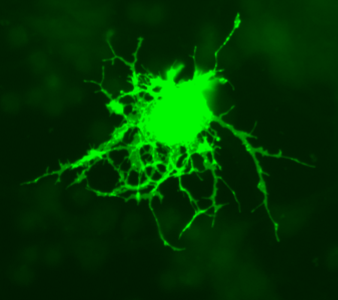
Microglia
Microglia are specialized macrophages capable of phagocytosis that protect neurons of the central nervous system. Though not technically glia because they are derived from hemopoietic precursors rather than ectodermal tissue, they are commonly categorized as such because of their supportive role to neurons.
These cells comprise approximately 15% of the total cells of the central nervous system. They are found in all regions of the brain and spinal cord. Microglial cells are small relative to macroglial cells, with changing shapes and oblong nuclei. They are mobile within the brain and multiply when the brain is damaged. In the healthy central nervous system, microglia processes constantly sample all aspects of their environment (neurons, macroglia and blood vessels).
Macroglia
| Location | Name | Description |
| CNS | Astrocytes |
The most abundant type of glial cell, astrocytes (also called astroglia) have numerous projections that anchor neurons to their blood supply. They regulate the external chemical environment of neurons by removing excess ions, notably potassium, and recycling neurotransmitters released during synaptic transmission. The current theory suggests that astrocytes may be the predominant “building blocks” of the blood-brain barrier. Astrocytes may regulate vasoconstriction and vasodilation by producing substances such as arachidonic acid, whose metabolites are vasoactive. Astrocytes signal each other using calcium. The gap junctions (also known as electrical synapses) between astrocytes allow the messenger molecule IP3 to diffuse from one astrocyte to another. IP3 activates calcium channels on cellular organelles, releasing calcium into the cytoplasm. This calcium may stimulate the production of more IP3. The net effect is a calcium wave that propagates from cell to cell. Extracellular release of ATP, and consequent activation of purinergic receptors on other astrocytes, may also mediate calcium waves in some cases. There are generally two types of astrocytes, protoplasmic and fibrous, similar in function but distinct in morphology and distribution. Protoplasmic astrocytes have short, thick, highly branched processes and are typically found in gray matter. Fibrous astrocytes have long, thin, less branched processes and are more commonly found in white matter. |
| CNS | Oligodendrocytes |
Oligodendrocytes are cells that coat axons in the central nervous system (CNS) with their cell membrane, called myelin, producing the so-called myelin sheath. The myelin sheath provides insulation to the axon that allows electrical signals to propagate more efficiently. |
| CNS | Ependymal cells |
Ependymal cells, also named ependymocytes, line the cavities of the CNS and make up the walls of the ventricles. These cells create and secrete cerebrospinal fluid(CSF) and beat their cilia to help circulate that CSF. |
| CNS | Radial glia |
Radial glia cells arise from neuroepithelial cells after the onset of neurogenesis. Their differentiation abilities are more restricted than those of neuroepithelial cells. In the developing nervous system, radial glia function both as neuronal progenitors and as a scaffold upon which newborn neurons migrate. In the mature brain, the cerebellum and retina retain characteristic radial glial cells. In the cerebellum, these are Bergmann glia, which regulate synaptic plasticity. In the retina, the radial Müller cell is the principal glial cell, and participates in a bidirectional communication with neurons. |
| PNS | Schwann cells |
Similar in function to oligodendrocytes, Schwann cells provide myelination to axons in the peripheral nervous system (PNS). They also have phagocytotic activity and clear cellular debris that allows for regrowth of PNS neurons. |
| PNS | Satellite cells |
Satellite cells are small cells that line the exterior surface of PNS neurons and help regulate the external chemical environment. |
Capacity to divide
Glia retain the ability to undergo cell division in adulthood, while most neurons cannot. The view is based on the general deficiency of the mature nervous system in replacing neurons after an insult or injury, such as a stroke or trauma, while very often there is a profound proliferation of glia, or gliosis near or at the site of damage. However, detailed studies found no evidence that ‘mature’ glia, such as astrocytes or oligodendrocytes, retain the ability of mitosis. Only the resident oligodendrocyte precursor cells seem to keep this ability after the nervous system matures. On the other hand, there are a few regions in the mature nervous system, such as the dentate gyrus of the hippocampus and the subventricular zone, where generation of new neurons can be observed.
Embryological development
Most glia are derived from ectodermal tissue of the developing embryo, particularly the neural tube and crest. The exception is microglia, which are derived from hemopoietic stem cells. In the adult, microglia are largely a self-renewing population and are distinct from macrophages and monocytes which infiltrate the injured and diseased CNS.
In the central nervous system, glia develop from the ventricular zone of the neural tube. These glia include the oligodendrocytes, ependymal cells, and astrocytes. In the peripheral nervous system, glia derive from the neural crest. These PNS glia include Schwann cells in nerves and satellite cells in ganglia.
History
Glia were discovered in 1856 by the pathologist Rudolf Virchow in his search for a ‘connective tissue’ in the brain.
The human brain contains about ten times more glial cells than neurons. [1] Following its discovery in the late 19th century, this fact underwent significant media distortion, emerging as the famous myth claiming that “we are using only 10% of our brain”. The role of glial cells as managers of communications in the synapse gap, thus modifying learning pace, has been discovered only very recently (2004).
Additional images
-
 Oligodendrocyte
 -
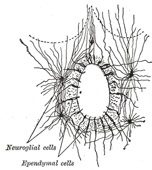 Section of central canal of medulla spinalis, showing ependymal and neuroglial cells.
 -
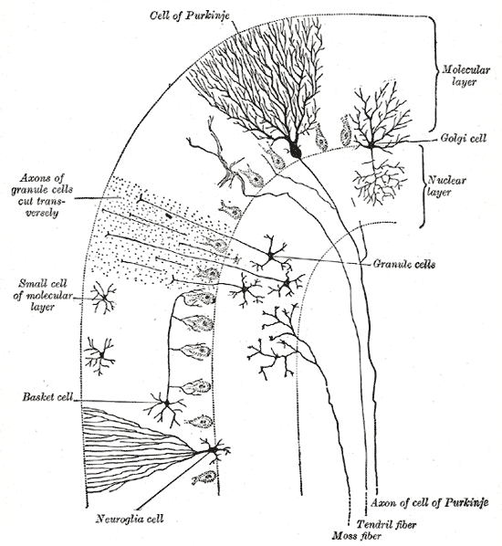 Transverse section of a cerebellar folium.


References
External links
- Role of glia in synapse development
- Overstreet L (2005). “Quantal transmission: not just for neurons”. Trends Neurosci. 28 (2): 59–62. PMID 15667925. article
- Peters A (2004). “A fourth type of neuroglial cell in the adult central nervous system”. J Neurocytol. 33 (3): 345–57. PMID 15475689.
- Volterra A, Steinhäuser C (2004). “Glial modulation of synaptic transmission in the hippocampus”. Glia. 47 (3): 249–57. PMID 15252814.
- Huang Y, Bergles D (2004). “Glutamate transporters bring competition to the synapse”. Curr Opin Neurobiol. 14 (3): 346–52. PMID 15194115.
- New Source of Replacement Brain Cells Found – glial cells can transform into other cell types and reproduce indefinitely — tricks once thought exclusive to stem cells.
- Artist ADSkyler(uses concepts of neuroscience and found inspiration from Glia)
de:Gliazelle eo:Glia ĉelo eu:Glia io:Glia celulo it:Cellula della glia he:תאי גליה nl:Gliacel sk:Ependýmová bunka fi:Gliasolu sv:Gliacell th:เซลล์เกลีย uk:Нейроглія Template:Jb1 Template:WH Template:WS
Lens cells
Lens cells
Germ cells
Germ cells
- Oogonium/Oocyte
- Spermatid
- Spermatocyte
- Spermatogonium cell (stem cell for spermatocyte)
- Spermatozoon
References
References
- Robert A. Freitas Jr., Nanomedicine, Volume I: Basic Capabilities, Landes Bioscience, Georgetown, TX, 1999
Looking for the patient version?
© 2026 MyEClinic – IFTM Institut für Telematik in der Medizin GmbH