
Mechanical ventilation
Editor-In-Chief: C. Michael Gibson, M.S., M.D. [1]; Associate Editor(s)-in-Chief: Ajay Gade MD[2]]Syed Hassan A. Kazmi BSc, MD [3]
Synonyms and keywords:: Intubation, Extubation, ventilator weaning, ventilator variables, vent, mechanically ventilated
Overview
Editor-In-Chief: C. Michael Gibson, M.S., M.D. [1] Associate Editor(s)-in-Chief: Ajay Gade MD[2]]Syed Hassan A. Kazmi BSc, MD [3]
Overview
In medicine, mechanical ventilation is a method to mechanically assist or replace spontaneous breathing when patients cannot do so on their own, and must be done so after invasive intubation with an endotracheal or tracheostomy tube through which air is directly delivered (in contrast to noninvasive ventilation). In many cases, mechanical ventilation is used in acute settings such as in the ICU for a short period of time during a serious illness. For some patients who have certain chronic illnesses that require long-term ventilation assistance, they are also able to do so at home or other nursing/rehabilitation institution with the help of respiratory therapists and physicians. The main form of mechanical ventilation currently is positive pressure ventilation, which works by increasing the pressure in the patient’s airway and thus forcing additional air into the lungs. This is in contrast to the more historically common negative pressure ventilators (for example, the “iron lung“) that create a negative pressure environment around the patient’s chest, thus sucking air into the lungs. Although often a life-saving technique, mechanical ventilation carries many potential complications including pneumothorax, airway injury, alveolar damage, and ventilator-associated pneumonia, among others. Accordingly it is generally weaned off or to minimal settings as soon as possible.
Historical Perspective
Vesalius was the first person to describe mechanical ventilation by inserting a reed or cane into the trachea of animals and then blowing into this tube. The iron lung, also known as the Drinker and Shaw tank, was developed in 1929 and was one of the first negative-pressure machines used for long-term ventilation.
Types of Ventilators
Mechanical ventilation may be classified into non-invasive and invasive mechanical ventilation. Non-invasive mechanical ventilation can be further sub-divided into continuous positive airway pressure breathing (CPAP), bilevel positive airway pressure breathing (BiPAP) and mask ventilation. Mechanical ventilators may also be classified based on the basic underlying mechanics of the device and the clinical condition in which it is used. Ventilation may be delivered via bag valve mask, continuous flow, transport ventilators, ICU ventilators, NICU ventilators and PAP ventilators
Indications for Use
Mechanical ventilation can be used in patients who have labored breathing and are unable to maintain adequate gaseous excange leading to hypoxemia and/or hypercapnia. Common clinical indications of mechanical ventilation include moderate to severe dyspnea, respiratory rate (RR) > 24-30/min, signs of increased breathing, accessory muscle use for breathing and abdominal paradox. It may also be used in patients who have inadequate arterial partial pressure of oxygen or critically low PaO2 (PaO2 < 70 mm Hg), hypercapnia PaCO2 > 45 mm Hg and PaO2/FiO2 < 200. Patients suffering from acute exacerbation of COPD, asthma/asthmatic attack, neuromuscular disease that prevents chest movement to allow gas exchange, central nervous system depression (CNS depression due to drugs, cardiac arrest, trauma), chest injury, chest malformation, acute and chronic respiratory failure, heart failure and ventilation-perfusion mismatch may also be candidates for mechanical ventilation.
Ventilator Variables
Ventilator variables modulate the oxygenation achieved. They can be adjusted according to the clinical condition of the patient and to achieve specific goals of management. The variables include fraction of inspired oxygen (FiO2), tidal volume (Vt), respiratory rate(f), positive end expiratory pressure (PEEP), inspiratory time, inspiratory flow rate, peak inspiratory pressure (PIP) and plateau pressure (Pplateau). Tailoring the ventilator settings can help achieve specific goals, for example, to improve oxygenation, option include increasing the FiO2 and PEEP and to improve ventilation, the tidal volume (Vt), inspiratory pressure and respiratory rate (f) may be increased (this follows the basic principle of minute ventilation = Tidal volume x respiratory rate).
Choosing Amongst Ventilator Modes
Choice of ventilator mode depends upon the clinical condition of the patient. Choice of ventilator mode can be tailored to achieve specific goals of management and set to achieve spontaneous breathing, volume-targeted ventilation, pressure-targeted ventilation, or some combination. In some conditions, for example in case of spontaneously breathing patient, the patient sets the respiratory rate and generates the desired flow rate. Different modes of ventilation include pressure support ventilation (PSV), continued mandatory ventilation (CMV) or assist control mode (AC), synchronous intermittent mandatory ventilation (SIMV), proportional assist ventilation (PAV), dual control mode, high frequency ventilation, pressure and volume targeted modes.
Initial Ventilator Settings
Initial ventilator settings should be modified and tailored according to the clinical condition of the patient and specific goals of management. Selection of ventilatory mode, sensitivity at flow trigger mode, tidal volume, rate, inspiratory flow, positive end expiratory pressure (PEEP), pressure limit, inspiratory time and fraction of inspired oxygen (FiO2) should be made according to the underlying etiology of hypoxemia/hypercapnia. Other factors for example, age of the patient, weight and height also play an important role in deciding the initial ventilatory settings. General rules that help physicians to choose the initial settings in a time-efficient manner include choosing a tidal volume of 12 mL per kg body weight delivered at a rate of 12 a minute (12-12 rule) in adults and adolescents. In infants and children without existing lung disease a tidal volume of 4-10 ml/kg may be delivered at a rate of 30-35 breaths per minute. With respiratory distress syndrome (RDS), a decreased tidal volume and increased respiratory rate sufficient to maintain pCO2between 45-55. Allowing higher pCO2 (sometimes called permissive hypercapnia) may help prevent ventilator induced lung injury. Ventilator triggered breaths may be initiated via either a pressure-triggered or a flow-triggered mechanism. Pressure triggered breaths are initiated when the ventilator senses a negative pressure (indicating that the patient is trying to initiate a breath). During the flow-triggered mechanism, a continuous flow is delivered and a ventilator-delivered breath is initiated when the return flow is less than the delivered flow.
Protocol
Candidacy for mechanical ventilation is based on specific criteria and clinical condition of the patient. Body weight of the patient and height also play important role in determining the optimal ventilator settings. Similar to initiation of mechanical ventilation there are specific criteria for weaning the patient off from the ventilator and doing a spontaneous breathing trial.
Complications
Complications of mechanical ventilation include, oxygen toxicity, ventilator associated pneumonia (VAP), laryngeal edema and ulceration, malnutrition, oversedation/delirium and ventilator induced lung injury..
Modification of Settings
In adults when 100% FiO2 is used initially, it is easy to calculate the next FiO2 to be used and easy to estimate the shunt fraction. When using 100% FiO2, the degree of shunting is estimated by subtracting the measured PaO2 (from an arterial blood gas) from 700 mmHg. For each difference of 100 mmHg, the shunt is 5%. A shunt of more than 25% should prompt a search for the cause of this hypoxemia, such as mainstem intubation or pneumothorax, and should be treated accordingly. Patients should have their ventilation considered for withdrawal if they are able to support their own ventilation and oxygenation, and this should be assessed continuously. Specific clinical, ventilator and oxygenation criteria should be met in order to do a trial of spontaneous breathing in mechanically ventilated patients.
Connection to Ventilator
There are various procedures and mechanical devices that provide protection against airway collapse, air leakage, and aspiration. Face mask, laryngeal mask airway, tracheal intubation, esophageal obturator airway, cricothyroidotomy and tracheostomy may be used to provide mechanical ventilation.
Terminology
References
Historical Perspective
Editor-In-Chief: C. Michael Gibson, M.S., M.D. [1] Associate Editor(s)-in-Chief: Vishnu Vardhan Serla M.B.B.S. [2]
Overview
Vesalius was the first person to describe mechanical ventilation by inserting a reed or cane into the trachea of animals and then blowing into this tube. The iron lung, also known as the Drinker and Shaw tank, was developed in 1929 and was one of the first negative-pressure machines used for long-term ventilation.
Historical Perspective
Vesalius was the first person to describe mechanical ventilation by inserting a reed or cane into the trachea of animals and then blowing into this tube.[1]
Negative Pressure Machines
- The iron lung, also known as the Drinker and Shaw tank, was developed in 1929 and was one of the first negative-pressure machines used for long-term ventilation.
- It was refined and used in the 20th century largely as a result of the polio epidemic that struck the world in the 1950s.
- The machine is effectively a large elongated tank, which encases the patient up to the neck.
- The neck is sealed with a rubber gasket so that the patient’s face (and airway) are exposed to the room air.
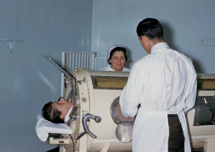
Iron Lung

- Gaseous exchange in the lungs takes place by process of simple diffusion, however, air must be drawn into the lungs to make it available for exchange.
- In spontaneous breathing, a negative pressure is created in the pleural cavity by the muscles of respiration, and the resulting gradient between the atmospheric pressure and the pressure inside the thorax generates air flow.
- In the iron lung, vacuum is created inside a tank by pumping out the air, thus generating a pressure gradient to allow flow of air.
- After the air is drawn into the lungs via negative tank pressure, the vacuum is released leading to equalization of pressure inside the tank and the ambient air pressure. This leads the air to exit the lungs due to natural elastic recoil of the lung and thoracic cage.
- Due to the vacuum created in the tank, the abdomen also expands along with the chest cavity leading to decreased venous return and pooling of blood in the extremities.
- The patients can talk and eat normally and can see the world through a well-placed series of mirrors.
- Some could remain in these iron lungs for years at a time quite successfully.
- Today, negative pressure mechanical ventilators are still in use, notably with the Polio Wing Hospitals in England such as St Thomas’ (by Westminster in London) and the John Radcliffe in Oxford.
- The prominent device used is a smaller device known as the cuirass.
- A cuirass is a shell-like unit, creating negative pressure only to the chest using a combination of a fitting shell and a soft bladder. Its main use is in patients with neuromuscular disorders who have some residual muscular function.
- However, it was prone to falling off and caused severe chafing and skin damage and was not used as a long-term device.
- In recent years this device has re-surfaced as a modern polycarbonate shell with multiple seals and a high-pressure oscillation pump in order to carry out biphasic cuirass ventilation.
Positive Pressure Machines
- The design of the modern positive-pressure ventilators was mainly based on technical developments by the military during World War II to supply oxygen to fighter pilots at high altitude.
- Such ventilators replaced the iron lungs as safe endotracheal tubes with high volume/low pressure cuffs were developed.

Mechanical ventilator - The popularity of positive-pressure ventilators rose during the polio epidemic in the 1950s in Scandinavia and the United States.
- Positive pressure through the manual supply of 50% oxygen through a tracheostomy tube led to a reduced mortality rate among patients with polio and respiratory paralysis.
- However, because of the sheer amount of manpower required for such intervention, positive-pressure ventilators became increasingly popular.

- Positive-pressure ventilators work by increasing the patient’s airway pressure through an endotracheal or tracheostomy tube. The positive pressure allows air to flow into the airway until the ventilator breath is terminated.
- Subsequently, the airway pressure drops to zero, and the elastic recoil of the chest wall and lungs push the tidal volume out through passive exhalation.
References
- ↑ Chamberlain D (2003) “Never quite there: A tale of resuscitation medicine” Clinical Medicine, Journal of the Royal College of Physicians’ 3 6:573-577
Types of Ventilators
Editor-In-Chief: C. Michael Gibson, M.S., M.D. [1] Associate Editor(s)-in-Chief: Vishnu Vardhan Serla M.B.B.S. [2]Syed Hassan A. Kazmi BSc, MD [3]
Overview
Mechanical ventilation may be classified into non-invasive and invasive mechanical ventilation. Non-invasive mechanical ventilation can be further sub-divided into continuous positive airway pressure breathing (CPAP), bilevel positive airway pressure breathing (BiPAP) and mask ventilation. Mechanical ventilators may also be classified based on the basic underlying mechanics of the device and the clinical condition in which it is used. Ventilation may be delivered via bag valve mask, continuous flow, transport ventilators, ICU ventilators, NICU ventilators and PAP ventilators.
Classification
Mechanical ventilation may be classified into the following types:
1. Non-invasive mechanical ventilation
Non-invasive mechanical ventilation may be further classified into:[1][2][3][4][5]
(a) Continuous positive airway pressure (CPAP)
- Equivalent to positive end expiratory pressure (PEEP)
- Patient breathes spontaneously at own rate
- Ventilator maintains a constant positive airway pressure throughout the respiratory cycle
- No limit of oxygen delivered (i.e. can give high flow oxygen FiO2 = 1)
- Used if primary problem is hypoxemia
(b) Bilevel positive airway pressure (BiPAP)
- Equivalent to positive end expiratory pressure (PEEP) + Pressure support ventilation (PSV)
- Able to set both inspiratory (usually 8-10 cm H2O) and expiratory pressure (usually < 5 cm H2O)
- Oxygen delivery limited
- Used if primary problem is hypovantilation
(c) Mask ventilation
- Tight-fitting mask connecting patient to a standard ventilator can receive pressure support of 20-30 cm H2O, PEEP of 10 cm H2O, FiO2 of 1.0
- Used for short-term reversible process (< 24 hours)
2. Invasive mechanical ventilation
Types of Ventilators

Ventilation can be delivered via:
- Hand-controlled ventilation such as:
- Bag valve mask
- Continuous-flow or anaesthesia (T-piece) bag
- Types of mechanical ventilators include:
- Transport ventilators:
- These ventilators are small and more rugged
- These are powered pneumatically, or via DC or AC power sources.
- ICU ventilators:
- These ventilators are larger and usually run on AC power.
- This style of ventilator often provides greater control of a wide variety of ventilation parameters such as inspiratory rise time.
- Many ICU ventilators also use graphics to provide visuals of each breath.
- NICU ventilators:
- These are a specialized subset of ICU ventilators which are designed to deliver the smaller, more precise volumes and pressures required to ventilate these patients.
- These are used specially for preterm babies.
- PAP ventilators: These ventilators are specifically designed for non-invasive ventilation at home for sleep apnea.
- Transport ventilators:
References
- ↑ Ball L, Dameri M, Pelosi P (2015). “Modes of mechanical ventilation for the operating room”. Best Pract Res Clin Anaesthesiol. 29 (3): 285–99. doi:10.1016/j.bpa.2015.08.003. PMID 26643095.
- ↑ Kacmarek RM, Pirrone M, Berra L (2015). “Assisted mechanical ventilation: the future is now!”. BMC Anesthesiol. 15: 110. doi:10.1186/s12871-015-0092-y. PMC 4517541. PMID 26215886.
- ↑ Kübler A, Maciejewski D, Adamik B, Kaczorowska M (2013). “Mechanical ventilation in ICUs in Poland: a multi-center point-prevalence study”. Med. Sci. Monit. 19: 424–9. doi:10.12659/MSM.883930. PMC 3673926. PMID 23727991.
- ↑ Muhlethaler V, Malcolm G (2014). “Mechanical ventilation in the newborn; a simplified approach. Part 2: High-frequency ventilation”. J Paediatr Child Health. 50 (10): E10–3. doi:10.1111/j.1440-1754.2010.01873.x. PMID 20977521.
- ↑ Yoshida T, Torsani V, Gomes S, De Santis RR, Beraldo MA, Costa EL, Tucci MR, Zin WA, Kavanagh BP, Amato MB (2013). “Spontaneous effort causes occult pendelluft during mechanical ventilation”. Am. J. Respir. Crit. Care Med. 188 (12): 1420–7. doi:10.1164/rccm.201303-0539OC. PMID 24199628.
Indications for Use
Editor-In-Chief: C. Michael Gibson, M.S., M.D. [1] Associate Editor(s)-in-Chief: Vishnu Vardhan Serla M.B.B.S. [2]Syed Hassan A. Kazmi BSc, MD [3]
Overview
Mechanical ventilation can be used in patients who have labored breathing and are unable to maintain adequate gaseous excange leading to hypoxemia and/or hypercapnia. Common clinical indications of mechanical ventilation include moderate to severe dyspnea, respiratory rate (RR) > 24-30/min, signs of increased breathing, accessory muscle use for breathing and abdominal paradox. It may also be used in patients who have inadequate arterial partial pressure of oxygen or critically low PaO2 (PaO2 < 70 mm Hg), hypercapnia PaCO2 > 45 mm Hg and PaO2/FiO2 < 200. Patients suffering from acute exacerbation of COPD, asthma/asthmatic attack, neuromuscular disease that prevents chest movement to allow gas exchange, central nervous system depression (CNS depression due to drugs, cardiac arrest, trauma), chest injury, chest malformation, acute and chronic respiratory failure, heart failure and ventilation-perfusion mismatch may also be candidates for mechanical ventilation.
Indications for Use
The indications of the mechanical ventilation is as follows:[1][2][3][4][5][6][7]
- The three most common indications for mechanical ventilation:
- Inadequate oxygenation
- Inadequate ventilation
- Inability to protect the airway
Other indications for mechanical ventilation include the following:
- Bradypnea
- Tachypnea (>30 breaths/minute)
- Apnea with respiratory arrest including cases from intoxication
- Acute respiratory distress syndrome
- Vital capacity less than 15 ml/kg
- Minute ventilation greater than 10 Lts/min
- Reduced respiratory drive
- Abnormalities of the chest wall
- Respiratory muscle fatigue
- Intrapulmonary shunt
- V/Q mismatch (ventilation-perfusion)
- Decreased Functional Residual Capacity
- Arterial partial pressure of oxygen (PaO2) with a supplemental fraction of inspired oxygen (FIO2) of less than 55 mm Hg
- Alveolar-arterial gradient of oxygen tension (A-a DO2) with 100% oxygenation of greater than 450 mm Hg
- Coma
- Hypotension due to sepsis, shock, CHF
- Acute partial pressure of carbon dioxide (PaCO2) greater than 50 mm Hg with an arterial pH less than 7.25
- Chronic obstructive pulmonary disease (COPD)
- Acute respiratory acidosis with
- Partial pressure of carbon dioxide (pCO2) > 50 mmHg
- pH < 7.25, which may be due to paralysis of the diaphragm due to
- Guillain-Barré syndrome
- Myasthenia Gravis
- spinal cord injury
- The effect of anaesthetic and muscle relaxants
- Increased work of breathing as evidenced by significant tachypnea, retractions, and other physical signs of respiratory distress
- Hypoxemia with arterial partial pressure of oxygen (PaO2) with supplemental fraction of inspired oxygen (FiO2) < 55 mm Hg
- Neuromuscular disease
References
- ↑ Tung A (1997). “Indications for mechanical ventilation”. Int Anesthesiol Clin. 35 (1): 1–17. PMID 9113518.
- ↑ Kreppein U, Litterst P, Westhoff M (2016). “[Hypercapnic respiratory failure. Pathophysiology, indications for mechanical ventilation and management]”. Med Klin Intensivmed Notfmed (in German). 111 (3): 196–201. doi:10.1007/s00063-016-0143-2. PMID 26902369.
- ↑ Strøm T, Rian O, Toft P (February 2012). “[Fewer indications for sedation in mechanical ventilation therapy]”. Ugeskr. Laeg. (in Danish). 174 (7): 406–9. PMID 22331041.
- ↑ Simonds AK (November 2016). “Home Mechanical Ventilation: An Overview”. Ann Am Thorac Soc. 13 (11): 2035–2044. doi:10.1513/AnnalsATS.201606-454FR. PMID 27560387.
- ↑ Boldrini R, Fasano L, Nava S (February 2012). “Noninvasive mechanical ventilation”. Curr Opin Crit Care. 18 (1): 48–53. doi:10.1097/MCC.0b013e32834ebd71. PMID 22186215.
- ↑ Cohen CA, Zagelbaum G, Gross D, Roussos C, Macklem PT (September 1982). “Clinical manifestations of inspiratory muscle fatigue”. Am. J. Med. 73 (3): 308–16. PMID 6812417.
- ↑ Slutsky AS (December 1993). “Mechanical ventilation. American College of Chest Physicians’ Consensus Conference”. Chest. 104 (6): 1833–59. PMID 8252973.
Modes of Ventilation
Editor-In-Chief: C. Michael Gibson, M.S., M.D. [1] Associate Editor(s)-in-Chief: Vishnu Vardhan Serla M.B.B.S. [2]
References
Choosing Amongst Ventilator Modes
Editor-In-Chief: C. Michael Gibson, M.S., M.D. [1] Associate Editor(s)-in-Chief: Syed Hassan A. Kazmi BSc, MD [2]
Overview
Choice of ventilator mode depends upon the clinical condition of the patient. Choice of ventilator mode can be tailored to achieve specific goals of management and set to achieve spontaneous breathing, volume-targeted ventilation, pressure-targeted ventilation, or some combination. In some conditions, for example in case of spontaneously breathing patient, the patient sets the respiratory rate and generates the desired flow rate. Different modes of ventilation include pressure support ventilation (PSV), continued mandatory ventilation (CMV) or assist control mode (AC), synchronous intermittent mandatory ventilation (SIMV), proportional assist ventilation (PAV), dual control mode, high frequency ventilation, pressure and volume targeted modes.
Choosing Amongst Ventilator Modes
Choice of ventilator mode depends upon the clinical condition of the patient. Choice of ventilator mode can be tailored to achieve specific goals of management and set to achieve spontaneous breathing, volume-targeted ventilation, pressure-targeted ventilation, or some combination. In some conditions, for example in case of spontaneously breathing patient, the patient sets the respiratory rate and generates the desired flow rate.[1][2][3] The following are the various ventilator modes and their features:[4][5][6][7][8][9]
Pressure support ventilation (PSV)
- This mode supports patient initiated breaths with a set inspiratory pressure and positive end expiratory pressure (PEEP)
- Mode of partial ventilatory support because there is no set rate
- The clinician sets the FiO2 and PEEP. The patient sets the respiratory rate and generates their desired flow rate.
- After the optimal flow is achieved, the applied pressure can be turned off.
- The volume given depends upon the patient’s effort and lung/chest wall compliance
- There is no minimal rate
Continued mandatory ventilation (CMV) or assist control (AC)
- Ventilator delivers a minimum number of supported breaths
- Mode of fully assisted ventilatory support
- Additional patient initiated breaths trigger fully assisted vent breaths. Therefore, vent-triggered breaths are identical to patient triggered breaths
- May be pressure targeted or volume targeted
- If patient develops tachypnea, may lead to respiratory alkalosis, breath stacking and auto PEEP
Synchronous intermittent mandatory ventilation (SIMV)
- Ventilator develops a minimum number of supported breaths
- Tidal volume of additional breaths is determined by the patients effort
- This strategy also supplies inspiratory pressure during spontaneous breaths (similar to pressure support ventilation)
- May be used to wean off patient after extubation (maybe combined with pressure support ventilation- PSV)
Proportional assist ventilation (PAV)
- Delivers variable pressure to achieve targeted percentage of work of breathing
Dual control modes
- Combination of volume and pressure targeted ventilation
- Use a closed loop ventilator logic
- Automatically alter control variables, either breath-to-breath or within a breath, to ensure a minimum tidal volume or minute-ventilation
High frequency ventilation
- Helps to achieve optimum gas exchange when using high respiratory rates (RR) with tidal volumes (Vt) lesser than the anatomical dead space
- Used in neonatal respiratory failure
- May be used in cases of acute lung injury (ALI) or acute respiratory distress syndrome (ARDS)
Volume targeted modes
- Both assist control (AC) and syncronous intermittent support ventilation (SIMV) can be volume controlled
- Ventilator delivers a set tidal volume (Vt)
- Pressures depend on lung compliance
Advantages
- Safe in cases of acute lung injury (ALI) and acute respiratory distress syndrome (ARDS)
- Ideal initial ventilator setting
- Easy to measure mechanics (PIP, Pplat, airway resistance, compliance)
Pressure targeted modes
- Ventilator delivers a fixed inspiratory pressure regardless of Vt
- Vt depends on lung/chest wall compliance
Advantages
- May increase patient comfort (PSV) requiring less sedation
Proportional assist ventilation plus and proportional pressure support
References
- ↑ “Treatment of severe cardiogenic pulmonary edema with continuous positive airway pressure delivered by face mask. – PubMed – NCBI”.
- ↑ “How is mechanical ventilation employed in the intensive care unit? An international utilization review. – PubMed – NCBI”.
- ↑ “Characteristics and outcomes in adult patients receiving mechanical ventilation: a 28-day international study. – PubMed – NCBI”.
- ↑ Singh PM, Borle A, Trikha A (July 2014). “Newer nonconventional modes of mechanical ventilation”. J Emerg Trauma Shock. 7 (3): 222–7. doi:10.4103/0974-2700.136869. PMC 4126124. PMID 25114434.
- ↑ Haas CF, Bauser KA (2012). “Advanced ventilator modes and techniques”. Crit Care Nurs Q. 35 (1): 27–38. doi:10.1097/CNQ.0b013e31823b2670. PMID 22157490.
- ↑ Rose L (May 2010). “Clinical application of ventilator modes: Ventilatory strategies for lung protection”. Aust Crit Care. 23 (2): 71–80. doi:10.1016/j.aucc.2010.03.003. PMID 20378369.
- ↑ Koh SO (April 2007). “Mode of mechanical ventilation: volume controlled mode”. Crit Care Clin. 23 (2): 161–7, viii. doi:10.1016/j.ccc.2006.11.014. PMID 17368163.
- ↑ Hess D (December 2001). “Ventilator modes used in weaning”. Chest. 120 (6 Suppl): 474S–6S. PMID 11742968.
- ↑ Botz GH, Sladen RN (1997). “Conventional modes of mechanical ventilation”. Int Anesthesiol Clin. 35 (1): 19–27. PMID 9113519.
Initial Ventilator Settings
Editor-In-Chief: C. Michael Gibson, M.S., M.D. [1] Associate Editor(s)-in-Chief: Vishnu Vardhan Serla M.B.B.S. [2]
Overview
Initial ventilator settings should be modified and tailored according to the clinical condition of the patient and specific goals of management. Selection of ventilatory mode, sensitivity at flow trigger mode, tidal volume, rate, inspiratory flow, positive end expiratory pressure (PEEP), pressure limit, inspiratory time and fraction of inspired oxygen (FiO2) should be made according to the underlying etiology of hypoxemia/hypercapnia. Other factors for example, age of the patient, weight and height also play an important role in deciding the initial ventilatory settings. General rules that help physicians to choose the initial settings in a time-efficient manner include choosing a tidal volume of 12 mL per kg body weight delivered at a rate of 12 a minute (12-12 rule) in adults and adolescents. In infants and children without existing lung disease a tidal volume of 4-10 ml/kg may be delivered at a rate of 30-35 breaths per minute. With respiratory distress syndrome (RDS), a decreased tidal volume and increased respiratory rate sufficient to maintain pCO2 between 45-55. Allowing higher pCO2 (sometimes called permissive hypercapnia) may help prevent ventilator induced lung injury. Ventilator triggered breaths may be initiated via either a pressure-triggered or a flow-triggered mechanism. Pressure triggered breaths are initiated when the ventilator senses a negative pressure (indicating that the patient is trying to initiate a breath). During the flow-triggered mechanism, a continuous flow is delivered and a ventilator-delivered breath is initiated when the return flow is less than the delivered flow.
Initial Ventilator Settings
The following are general guidelines that may need to be modified for the individual patient.[1][2][3][4][5][6][7][8][9][10]
Tidal Volume, Rate, and Pressures
- Adult patients and older children
- Without existing lung disease — a tidal volume of 12 mL per kg body weight is set to be delivered at a rate of 12 a minute (12-12 rule).
- With COPD — a reduced tidal volume of 10 ml/kg is to be delivered 10 times a minute to prevent overinflation and hyperventilation (10-10 rule).
- With acute respiratory distress syndrome (ARDS) — an even more reduced tidal volume of 6-8 mL/kg is used with a rate of 10-12/minute. This reduced tidal volume allows for minimal volutrauma but may result in an elevated pCO2 (due to the relative decreased oxygen delivered) but this elevation does not need to be corrected (termed permissive hypercapnia)
- Infants and younger children
- Without existing lung disease — a tidal volume of 4-10 ml/kg to be delivered at a rate of 30-35 breaths per minute
- With RDS — decrease tidal volume and increase respiratory rate sufficient to maintain pCO2 between 45-55. Allowing higher pCO2 (sometimes called permissive hypercapnia) may help prevent ventilator induced lung injury
- As the amount of tidal volume increases, the pressure required to administer that volume is increased.
- This pressure is known as the peak airway pressure. If the peak airway pressure is persistently above 45 cmH2O for adults, the risk of barotrauma is increased and efforts should be made to try to reduce the peak airway pressure.
- In infants and children, it is unclear what level of peak pressure may cause damage. In general, keeping peak pressures below 30 is desirable.
- Monitoring for barotrauma can also involve measuring the plateau pressure, which is the pressure after the delivery of the tidal volume but before the patient is allowed to exhale.
- Normal breathing pattern involves inspiration, then expiration. The ventilator is programmed so that after delivery of the tidal volume (inspiration), the patient is not allowed to exhale for a half a second.
- Therefore, pressure must be maintained in order to prevent exhalation, and this pressure is the plateau pressure. Barotrauma is minimized when the plateau pressure is maintained < 30-35 cmH2O.
Sighs
- An adult patient breathing spontaneously will usually sigh about 6-8 times/hr to prevent microatelectasis, and this has led some to propose that ventilators should deliver 1.5-2 times the amount of the preset tidal volume 6-8 times/hr to account for the sighs.
- However, such high quantity of volume delivery requires very high peak pressure that predisposes to barotrauma.
- Currently, accounting for sighs is not recommended if the patient is receiving 10-12 mL/kg or is on PEEP.
- If the tidal volume used is lower, the sigh adjustment can be used, as long as the peak and plateau pressures are acceptable.
- Sighs are not generally used with ventilation of infants and young children.
Initial FiO2
- Because the mechanical ventilator is responsible for assisting in a patient’s breathing, it must then also be able to deliver an adequate amount of oxygen in each breath.
- The FiO2 stands for fraction of inspired oxygen, which means the percent of oxygen in each breath that is inspired. (Note that normal room air has ~21% oxygen content).
- In adult patients who can tolerate higher levels of oxygen for a period of time, the initial FiO2 may be set at 100% until arterial blood gases can document adequate oxygenation.
- An FiO2 of 100% for an extended period of time can be dangerous, but it can protect against hypoxemia from unexpected intubation problems. For infants, and especially in premature infants, avoiding high levels of FiO2 (>60%) is important.
Positive End-Expiratory Pressure (PEEP)
- PEEP is an adjuvant to the mode ventilation used in cases where the functional residual capacity (FRC) is reduced.
- At the end of expiration, the PEEP exerts pressure to oppose passive emptying of the lung and to keep the airway pressure above the atmospheric pressure.
- The presence of PEEP opens up collapse or unstable alveoli and increases the FRC and surface area for gas exchange, thus reducing the size of the shunt.
- Thus, if a large shunt is found to exist based on the estimation from 100% FiO2, then PEEP can be considered and the FiO2 can be lowered (< 60%) to still maintain an adequate PaO2, thus reducing the risk of oxygen toxicity.
- In addition to treating a shunt, PEEP is also therapeutic in decreasing the work of breathing. In pulmonary physiology, compliance is a measure of the “stiffness” of the lung and chest wall.
- The mathematical formula for compliance (C) = change in volume/change in pressure. Therefore, a higher compliance means that only small increases in pressure can lead to large increases in volume, which means the work of breathing is reduced.
- As the FRC increases with PEEP, the compliance also increases, since the partially inflated lung takes less energy to inflate further.
Indications
- PEEP is a cardio-depressant and can cause severe hemodynamic consequences through decreasing venous return to the right heart and decreasing right ventricular outflow. As such, it should be judiciously used and is indicated in two circumstances.
- If used, PEEP is usually set with the minimal positive pressure to maintain an adequate PaO2 with a safe FiO2.
- As PEEP increase intrathoracic pressure, there can be a resulting decrease in venous return and decrease in cardiac output. A PEEP of less than 10 cmH2O is usually safe if intravascular volume depletion is absent.
- Older literature recommended routine placement of a Swan-Ganz catheter if the amount of PEEP used is greater than 10 cmH2 for hemodynamic monitoring.
- More recent literature has failed to find outcome benefits with routine PA catheterization when compared to simple central venous pressure monitoring.[11]
- If cardiac output measurement is required, minimally invasive techniques, such as esophageal Doppler monitoring or arterial waveform contour monitoring may be sufficient alternatives.
- PEEP should be withdrawn from a patient until adequate PaO2 can be maintained with a FiO2 < 40%. When withdrawing, it is decreased through 1-2 cmH2O decrements while monitoring hemoglobin-oxygen saturation.
- Any unacceptable hemoglobin-oxygen saturation should prompt re-institution of the last PEEP level that maintained good saturation.
Positioning
Prone (face down) positioning has been used in patients with ARDS and severe hypoxemia. It improves FRC, drainage of secretions, and ventilation-perfusion matching (efficiency of gas exchange). It may improve oxygenation in > 50% of patients, but no survival benefit has been documented.
Sedation
Most patients receive sedation through a continuous infusion or scheduled dosing to help with anxiety or psychological stress. Daily interruption of sedation is commonly helpful to the patient for reorientation and appropriate weaning.
Prophylaxis
- To protect against ventilator-associated pneumonia, patient’s bed is often elevated to about 30° and Histamine-2 receptor blocker or proton pump inhibitors may be used.
- Deep vein thrombosis prophylaxis with heparin or sequential compression device is important in older children and adults.
References
- ↑ Vallee F, et al. Stroke output variations calculated by esophageal Doppler is a reliable predictor of fluid response. Intensive Care Med. 2005 Oct;31(10):1388-93. Epub 2005 August 19. PMID: 16132887
- ↑ Uchino S, et al. Pulmonary artery catheter versus pulse contour analysis: a prospective epidemiological study. Crit Care. 2006 December 14;10(6):R174 [Epub ahead of print] PMID: 17169160
- ↑ Bagga S, Paluzzi DE, Chen CY, Riggio JM, Nagaraja M, Marik PE, Baram M (August 2014). “Better ventilator settings using a computerized clinical tool”. Respir Care. 59 (8): 1172–7. doi:10.4187/respcare.02223. PMID 24327745.
- ↑ Fortis S, Florindez J, Balasingham S, De Aguirre M, Amoateng-Adjepong Y, Manthous CA (July 2015). “Ventilator Settings Can Substantially Impact Patients’ Comfort”. J Intensive Care Med. 30 (5): 286–91. doi:10.1177/0885066613519574. PMID 24446238.
- ↑ Akbulut FP, Akkur E, Akan A, Yarman BS (January 2014). “A decision support system to determine optimal ventilator settings”. BMC Med Inform Decis Mak. 14: 3. doi:10.1186/1472-6947-14-3. PMC 3996182. PMID 24410995.
- ↑ Falaize L, Leroux K, Prigent H, Louis B, Khirani S, Orlikowski D, Fauroux B, Lofaso F (July 2014). “Battery life of portable home ventilators: effects of ventilator settings”. Respir Care. 59 (7): 1048–52. doi:10.4187/respcare.02711. PMID 24149669.
- ↑ Kilickaya O, Gajic O (March 2013). “Initial ventilator settings for critically ill patients”. Crit Care. 17 (2): 123. doi:10.1186/cc12516. PMC 3672640. PMID 23510269.
- ↑ Wilcox SR, Richards JB, Fisher DF, Sankoff J, Seigel TA (August 2016). “Initial mechanical ventilator settings and lung protective ventilation in the ED”. Am J Emerg Med. 34 (8): 1446–51. doi:10.1016/j.ajem.2016.04.027. PMID 27139256.
- ↑ Das A, Menon PP, Hardman JG, Bates DG (June 2013). “Optimization of mechanical ventilator settings for pulmonary disease states”. IEEE Trans Biomed Eng. 60 (6): 1599–607. doi:10.1109/TBME.2013.2239645. PMID 23322759.
- ↑ Rose L, Kenny L, Tait G, Mehta S (February 2014). “Ventilator settings and monitoring parameter targets for initiation of continuous mandatory ventilation: a questionnaire study”. J Crit Care. 29 (1): 123–7. doi:10.1016/j.jcrc.2013.10.018. PMID 24331947.
- ↑ Shah, MR et al Impact of the pulmonary artery catheter in critically ill patients: a meta-analysis of randomized clinical trials. JAMA. 2005 October 5;294(13):1664-70. PMID: 16204666
Protocol
Editor-In-Chief: C. Michael Gibson, M.S., M.D. [1] Associate Editor(s)-in-Chief: Vishnu Vardhan Serla M.B.B.S. [2]
Overview
Candidacy for mechanical ventilation is based on specific criteria and clinical condition of the patient. Body weight of the patient and height also play important role in determining the optimal ventilator settings. Similar to initiation of mechanical ventilation there are specific criteria for weaning the patient off from the ventilator and doing a spontaneous breathing trial.
Protocol
Inclusion Criteria
- PaO2 <= 300 ( corrected for altitude)
- Bilateral (patchy, diffuse or homogenous) infiltrates consistent with pulmonary edema
- No clinical evidence of left atrial hypertension
Ventilator Setup
- Calculate predicted body weight. It is calculated using the formula
- Males = 50 + 2.3[height(in inches)-60]
- Females = 45.5 + 2.3[height(in inches)-60]
- Selecting the ventilator mode
- Set ventilator settings to achieve initial Vt = 8 ml/kg of predicted body weight
- Reduce VT by 1 ml/kg at intervals ≤ 2 hours until VT = 6ml/kg.
- Set initial rate to approximate baseline minute ventilation (not > 35 breaths per minute).
- Adjust VT and respiratory rate to achieve pH and plateau pressure goals below.
Oxygenation Goal
- PaO2 55-80 mmHg or SpO2 88-95% is the goal.
- Use a minimum PEEP of 5 cm H2O.
- Consider use of incremental FiO2/PEEP combinations to achieve goal.
- Lower PEEP/Higher FiO2
| FiO2 | 0.3 | 0.4 | 0.4 | 0.5 | 0.5 | 0.6 | 0.7 | 0.7 | 0.7 | 0.8 | 0.9 | 0.9 | 0.9 | 1.0 |
| PEEP | 5 | 5 | 8 | 8 | 10 | 10 | 10 | 12 | 14 | 14 | 14 | 16 | 18 | 18-24 |
- Higher PEEP/Lower FiO2
| FiO2 | 0.3 | 0.3 | 0.3 | 0.3 | 0.3 | 0.4 | 0.4 | 0.5 | 0.5 | 0.5 – 0.8 | 0.8 | 0.9 | 1.0 | 1.0 |
| PEEP | 5 | 8 | 10 | 12 | 14 | 14 | 16 | 16 | 18 | 20 | 22 | 22 | 22 | 24 |
Plateau Pressure Goal
- Plateau pressure goal(Pplat) is <= 30 cm H2O
- Check Pplat every 4th hourly after change in PEEP ot VT
- If Pplat > 30 cm of H2O
- Decrease VT by 1ml/kg
- If Pplat < 25 cm of H2O and VT < 6ml/kg
- Increase VT by 1ml/kg unbtil Pplat > 25 cm H2O or VT = 6 ml/kg
- If Pplat < 30 cm and breath stacking or dys-synchrony occurs
- Increase VT in 1 ml/kg increments to 7 or 8 ml/kg, if Pplat remains <=30 cm of H2O
PH Goal
- pH should be maintained at 7.30 – 7.45
- If pH is less than 7.30 (acidosis)
a. Range of 7.15 – 7.30
- Increase respiratory rate until pH > 7.30
- Maximum rate can be 35 breaths/min
b. Less than 7.15
- Increase respiratory rate to 35
- VT can be increased in 1 ml/kg until pH >7.15 (Pplat target of 30 may be exceeded)
- Bicarbonate can be given
- If pH is more than 7.45 (alkalosis)
- Decrease the ventilation rate if possible
I:E Ratio Goal
Recommend that duration of inspiration be less than equal to duration of expiration.
Weaning
A spontaneous breathing trial has to be done daily when
- FiO2 ≤ 0.40 and PEEP ≤ 8.
- PEEP and FiO2 ≤ values of previous day.
- Patient has acceptable spontaneous breathing efforts. (May decrease vent rate by 50% for 5 minutes to detect effort.)
- Systolic BP ≥ 90 mm Hg without vasopressor support.
- No neuromuscular blocking agents or blockade.
Spontaneous Breathing Trial
If all above criteria are met and patient has been in the observed for at least 12 hours, initiate a trial of upto 120 minutes of spontaneous breathing with FiO2 < 0.5 and PEEP < 5
1. Place on T-piece, trach collar, or CPAP ≤ 5 cm H2O with PS < 5
2. Assess for tolerance as below for up to two hours.
a. SpO2 ≥ 90: and/or PaO2 ≥ 60 mm Hg b. Spontaneous VT ≥ 4 ml/kg predicted body weight c. RR ≤ 35/min d. pH ≥ 7.3 e. No respiratory distress (distress= 2 or more)
- HR > 120% of baseline
- Marked accessory muscle use
- Abdominal paradox
- Diaphoresis
- Marked dyspnea
3. If tolerated for at least 30 minutes, consider extubation.
4. If not tolerated resume pre-weaning settings.
References
- ↑ Loss SH, de Oliveira RP, Maccari JG, Savi A, Boniatti MM, Hetzel MP, Dallegrave DM, Balzano Pde C, Oliveira ES, Höher JA, Torelly AP, Teixeira C (2015). “The reality of patients requiring prolonged mechanical ventilation: a multicenter study”. Rev Bras Ter Intensiva. 27 (1): 26–35. doi:10.5935/0103-507X.20150006. PMC 4396894. PMID 25909310.
- ↑ Grebennikov VA, Kriakvina OA, Bolunova ES, Degtiareva MV (2013). “[Prognostic criteria of the premature infants weaning from mechanical ventilation during trigger ventilation]”. Anesteziol Reanimatol (in Russian) (1): 26–30. PMID 23808249.
- ↑ Valenzuela J, Araneda P, Cruces P (March 2014). “Weaning from mechanical ventilation in paediatrics. State of the art”. Arch. Bronconeumol. 50 (3): 105–12. doi:10.1016/j.arbres.2013.02.003. PMID 23542044.
- ↑ Al Ashry HS, Modrykamien AM (2014). “Humidification during mechanical ventilation in the adult patient”. Biomed Res Int. 2014: 715434. doi:10.1155/2014/715434. PMC 4096064. PMID 25089275.
- ↑ Wielenga JM, van den Hoogen A, van Zanten HA, Helder O, Bol B, Blackwood B (March 2016). “Protocolized versus non-protocolized weaning for reducing the duration of invasive mechanical ventilation in newborn infants”. Cochrane Database Syst Rev. 3: CD011106. doi:10.1002/14651858.CD011106.pub2. PMID 26998745.
- ↑ Toft P, Olsen HT, Jørgensen HK, Strøm T, Nibro HL, Oxlund J, Wian KA, Ytrebø LM, Kroken BA, Chew M (December 2014). “Non-sedation versus sedation with a daily wake-up trial in critically ill patients receiving mechanical ventilation (NONSEDA Trial): study protocol for a randomised controlled trial”. Trials. 15: 499. doi:10.1186/1745-6215-15-499. PMC 4307177. PMID 25528350.
Modification of Settings
Editor-In-Chief: C. Michael Gibson, M.S., M.D. [1] Associate Editor(s)-in-Chief: Vishnu Vardhan Serla M.B.B.S. [2] Syed Hassan A. Kazmi BSc, MD [3]
Overview
In adults when 100% FiO2 is used initially, it is easy to calculate the next FiO2 to be used and easy to estimate the shunt fraction. When using 100% FiO2, the degree of shunting is estimated by subtracting the measured PaO2 (from an arterial blood gas) from 700 mmHg. For each difference of 100 mmHg, the shunt is 5%. A shunt of more than 25% should prompt a search for the cause of this hypoxemia, such as mainstem intubation or pneumothorax, and should be treated accordingly. Patients should have their ventilation considered for withdrawal if they are able to support their own ventilation and oxygenation, and this should be assessed continuously. Specific clinical, ventilator and oxygenation criteria should be met in order to do a trial of spontaneous breathing in mechanically ventilated patients.
Modification of Settings
- Modification of ventilator settings:[1][2][3][4]
- In adults when 100% FiO2 is used initially, it is easy to calculate the next FiO2 to be used and easy to estimate the shunt fraction.
- The estimated shunt fraction refers to the amount of oxygen not being absorbed into the circulation.
- In normal physiology, gas exchange (oxygen/carbon dioxide) occurs at the level of the alveoli in the lungs.
- The existence of a shunt refers to any process that hinders this gas exchange, leading to wasted oxygen inspired and the flow of unoxygenated blood back to the left heart (which ultimately supplies the rest of the body with unoxygenated blood).
- When using 100% FiO2, the degree of shunting is estimated by subtracting the measured PaO2 (from an arterial blood gas) from 700 mmHg. For each difference of 100 mmHg, the shunt is 5%.
- A shunt of more than 25% should prompt a search for the cause of this hypoxemia, such as mainstem intubation or pneumothorax, and should be treated accordingly.
- If such complications are not present, other causes must be sought after, and PEEP should be used to treat this intrapulmonary shunt. Other such causes of a shunt include:
- Alveolar collapse from major atelectasis.
- Alveolar collection of material other than gas, such as pus from pneumonia, water and protein from acute respiratory distress syndrome, water from congestive heart failure, or blood from hemorrhage
When to Withdraw Mechanical Ventilation
- Withdrawal from mechanical ventilation (also known as weaning) should not be delayed unnecessarily, nor should it be done prematurely.
- Patients should have their ventilation considered for withdrawal if they are able to support their own ventilation and oxygenation, and this should be assessed continuously.
Clinical criteria
- Resolution of acute phase of disease
- Adequate cough reflex
- No excessive secretions
- Hemodynamic stability
Ventilator criteria
- PaCO2 < 50 mm Hg
- Vital capacity > 10 ml/Kg
- Spontaneous tidal volume > 5 ml/Kg
- Spontaneous respiratory rate (f) < 35/min
- f/Vt <100 breaths/ml/min
- Minute ventilation < 10 L with ABGs within normal limits
Oxygenation criteria
- PaO2 without PEEP > 60 mm Hg at FiO2 0.4
- PaO2 with PEEP (< 8 cm H2O) > 100 m Hg at FiO2 0.4
- SaO2 > 90% at FiO2 0.4
- PaO2/FiO2 greater than equal to 150
- P(A-a)O2 < 350 mm Hg at FiO2 1.0
Non-Invasive Ventilation (Non-Invasive Positive Pressure Ventilation or NIPPV)
- This refers to all modalities that assist ventilation without the use of an endotracheal tube.
- Non-invasive ventilation is primarily aimed at minimizing patient discomfort and the complications associated with invasive ventilation.
- It is often used in cardiac disease, exacerbations of chronic pulmonary disease, sleep apnea, and neuromuscular diseases.
- Non-invasive ventilation refers only to the patient interface and not the mode of ventilation used; modes may include spontaneous or control modes and may be either pressure or volume modes.
- Some commonly used modes of NIPPV include:
- Continuous positive airway pressure (CPAP)
- Bi-level Positive Airway Pressure (BIPAP) pressures alternate between Inspiratory Positive Airway Pressure (IPAP) and a lower Expiratory Positive Airway Pressure (EPAP), triggered by patient effort. On many such devices, backup rates may be set, which deliver IPAP pressures even if patients fail to initiate a breath.
- Intermittent positive pressure ventilation (IPPV) via mouthpiece or mask
References
- ↑ Bordes J, Erwan d, Savoie PH, Montcriol A, Goutorbe P, Kaiser E (September 2014). “FiO2 delivered by a turbine portable ventilator with an oxygen concentrator in an Austere environment”. J Emerg Med. 47 (3): 306–12. doi:10.1016/j.jemermed.2014.04.033. PMID 24950943. Vancouver style error: initials (help)
- ↑ d’Aranda E, Bordes J, Bourgeois B, Clay J, Esnault P, Cungi PJ, Goutorbe P, Kaiser E, Meaudre E (2016). “Fraction of Inspired Oxygen Delivered by Elisée™ 350 Turbine Transport Ventilator With a Portable Oxygen Concentrator in an Austere Environment”. J Spec Oper Med. 16 (3): 30–35. PMID 27734439.
- ↑ Magill SS, Rhodes B, Klompas M (August 2014). “Improving ventilator-associated event surveillance in the National Healthcare Safety Network and addressing knowledge gaps: update and review”. Curr. Opin. Infect. Dis. 27 (4): 394–400. doi:10.1097/QCO.0000000000000083. PMC 4638221. PMID 24945615.
- ↑ Mira JP, Brunet F, Belghith M, Soubrane O, Termignon JL, Renaud B, Hamy I, Monchi M, Deslande E, Fierobe L (January 1995). “Reduction of ventilator settings allowed by intravenous oxygenator (IVOX) in ARDS patients”. Intensive Care Med. 21 (1): 11–7. PMID 7560467.
Connection to Ventilators
Editor-In-Chief: C. Michael Gibson, M.S., M.D. [1] Associate Editor(s)-in-Chief: Vishnu Vardhan Serla M.B.B.S. [2]
Overview
There are various procedures and mechanical devices that provide protection against airway collapse, air leakage, and aspiration. Face mask, laryngeal mask airway, tracheal intubation, esophageal obturator airway, cricothyroidotomy and tracheostomy may be used to provide mechanical ventilation.
Connection to Ventilators
There are various procedures and mechanical devices that provide protection against airway collapse, air leakage, and aspiration:[1][2][3][4][5][5][6]
- Face mask – In resuscitation and for minor procedures under anesthesia, a face mask is often sufficient to achieve a seal against air leakage. Airway patency of the unconscious patient is maintained either by manipulation of the jaw or by the use of nasopharyngeal or oropharyngeal airway. These are designed to provide a passage of air to the pharynx through the nose or mouth, respectively. Poorly fitted masks often cause nasal bridge ulcers which is a problem for some patients. Face masks are also used for non-invasive ventilation in conscious patients. A face mask does not, however, provide protection against aspiration.
- Laryngeal mask airway – The laryngeal mask airway (LMA), causes less pain and coughing than a tracheal tube. However, unlike tracheal tubes it does not seal against aspiration, making careful individualised evaluation and patient selection mandatory.
- Tracheal intubation is often performed for mechanical ventilation of hours to weeks duration. A tube is inserted through the nose (nasotracheal intubation) or mouth (orotracheal intubation) and advanced into the trachea. In most cases tubes with inflatable cuffs are used for protection against leakage and aspiration. Intubation with a cuffed tube is thought to provide the best protection against aspiration. Tracheal tubes inevitably cause pain and coughing. Therefore, unless a patient is unconscious or anesthetized for other reasons, sedative drugs are usually given to provide tolerance of the tube. Other disadvantages of tracheal intubation include damage to the mucosal lining of the nasopharynx or oropharynx and subglottic stenosis.
- Esophageal obturator airway – commonly used by emergency medical technicians, if they are not authorized to intubate (the “esophageal airway” familiar to fans of the television series, Emergency!). It is a tube which is inserted into the esophagus, past the epiglottis. Once it is inserted, a bladder at the tip of the airway is inflated, to block (“obturate”) the esophagus, and air or oxygen is delivered through a series of holes in the side of the tube.
- Cricothyrotomy – Patients who require emergency airway management, in whom tracheal intubation has been unsuccessful, may require an airway inserted through a surgical opening in the cricothyroid membrane. This is similar to a tracheostomy but a cricothyrotomy is reserved for emergency access. [3]
- Tracheostomy – When patients require mechanical ventilation for several weeks a tracheostomy may provide the most suitable access to the patient’s trachea. A tracheostomy is a surgically created passage into the trachea. Tracheostomy tubes are well tolerated and often do not necessitate any use of sedative drugs. Tracheostomy tubes may be inserted early during treatment in patients with pre-existing severe respiratory disease, or in any patient who are expected to be difficult to wean from mechanical ventilation, i.e., patients who have little muscular reserve.
References
- ↑ Ferrone G, Cipriani F, Spinazzola G, Festa O, Arcangeli A, Proietti R, Antonelli M, Conti G, Costa R (September 2013). “A bench study of 2 ventilator circuits during helmet noninvasive ventilation”. Respir Care. 58 (9): 1474–81. doi:10.4187/respcare.02060. PMID 23431311.
- ↑ “Hazard Report. Medical vacuum system connection to ventilator breathing circuit may have contributed to patient’s death”. Health Devices. 38 (3): 90–1. March 2009. PMID 19580096.
- ↑ Boukhettala N, Porée T, Diot P, Vecellio L (April 2015). “In vitro performance of spacers for aerosol delivery during adult mechanical ventilation”. J Aerosol Med Pulm Drug Deliv. 28 (2): 130–6. doi:10.1089/jamp.2013.1091. PMID 25050644.
- ↑ Türköz A, Balcı ŞT, Gönen H, Çınar Ö, Özker E, Türköz R (2014). “The effects of different ventilator modes on cerebral tissue oxygen saturation in patients with bidirectional superior cavopulmonary connection”. Ann Card Anaesth. 17 (1): 10–5. doi:10.4103/0971-9784.124122. PMID 24401296.
- ↑ 5.0 5.1 Dai B, Kang J, Sun LF, Tan W, Zhao HW (April 2014). “Influence of exhalation valve and nebulizer position on albuterol delivery during noninvasive positive pressure ventilation”. J Aerosol Med Pulm Drug Deliv. 27 (2): 125–32. doi:10.1089/jamp.2012.1024. PMID 23668546.
- ↑ Hidalgo V, Giugliano-Jaramillo C, Pérez R, Cerpa F, Budini H, Cáceres D, Gutiérrez T, Molina J, Keymer J, Romero-Dapueto C (2015). “Noninvasive Mechanical Ventilation in Acute Respiratory Failure Patients: A Respiratory Therapist Perspective”. Open Respir Med J. 9: 120–6. doi:10.2174/1874306401509010120. PMC 4541452. PMID 26312104.
Terminology
Editor-In-Chief: C. Michael Gibson, M.S., M.D. [1] Associate Editor(s)-in-Chief: Vishnu Vardhan Serla M.B.B.S. [2]
Terminology [1]
Terminology used in the field of mechanical ventilation and respiratory support:
- ASB Assisted Spontaneous Breathing — , also ASV = Assisted Spontaneous Ventilation
- ASV Adaptive Support Ventilation — closed-loop mechanical respiration, a further development of MMV
- PSV Pressure Support Ventilation — supported spontaneous respiration, see also ASB
- APRV Airway Pressure Release Ventilation
- BIPAP Biphasic Positive Airway Pressure
- TNI Therapy with Nasal Insufflation — nasal High-Flow mechanical ventialtion for respiration support
- CPAP Continuous Positive Airway Pressure
- CPPV Continuous Positive Pressure Ventilation
- EPAP Expiratory Positive Airway Pressure
- CMV Continuous Mandatory Ventilation
- PCMV (P-CMV) Pressure Controlled Mandatory Ventilation
- VCMV (V-CMV) Volume Controlled Mandatory Ventilation
- PC Pressure Control — pressure controlled, fully mechanical ventilation
- PCV Pressure Controlled Ventilation — pressure controlled, fully mechanical ventilation
- VCV Volume Controlled Ventilation — volume controlled, fully mechanical ventilation
- S-CPPV Synchronized Continuous Positive Pressure Ventilation
- HFV High Frequency Ventilation
- LFPPV Low Frequency Positive Pressure Ventilation
- ILV Independent Lung Ventilation — separate sides positive pressure ventilation
- IPAP Inspiratory Positive Airway Pressure
- IPPV Intermittent Positive Pressure Ventilation
- S-IPPV Synchronized Intermittent Positive Pressure Ventilation
- (S)IMV (Synchronized) Intermittent Mandatory Ventilation
- MMV Mandatory Minute Volume
- IRV Inversed Ratio Ventilation — mechanical ventilation with switched respiration phases/time rate
- PEEP Positive End-Expiratory Pressure
- PNPV Positive Negative Pressure Ventilation — switching pressure mechanical ventilation
- ZAP Zero Airway Pressure — Spontaneous respiration under Atmospheric pressure
- PPS Proportional Pressure Support
- ATC Automatic Tube Compensation
References
Looking for the patient version?
© 2026 MyEClinic – IFTM Institut für Telematik in der Medizin GmbH