
Hypoplastic left heart syndrome pathophysiology
Editor-In-Chief: C. Michael Gibson, M.S., M.D. [1]; Associate Editor(s)-In-Chief: Priyamvada Singh, M.B.B.S.[2], Cafer Zorkun, M.D., Ph.D. [3], Keri Shafer, M.D. [4]; Assistant Editor(s)-In-Chief: Kristin Feeney, B.S.[5]
Overview
Overview
In patients with hypoplastic left heart syndrome, the left side of the heart is unable to send enough blood to the body. As a result, the right side of the heart must maintain the circulation for both the lungs and the body. The right ventricle can support the circulation to both the lungs and the body for a while, but this extra workload eventually causes the right side of the heart to fail.
Pathophysiology
Pathophysiology
The only possibility of survival is a connection between the right and the left side of the heart, or between the arteries and pulmonary arteries (the blood vessels that carry blood to the lungs).In babies with HLHS, the aorta and left ventricle are very small, and the aortic and mitral valves are either too small to allow sufficient blood flow or are atretic (closed) altogether. As blood returns from the lungs to the left atrium, it must pass through an atrial septal defect to the right side of the heart. In a healthy human, the left side of the heart receives oxygen-rich blood from the lungs and pumps it out to the rest of the body; with these structures underdeveloped, they cannot circulate blood to other organs, and the right ventricle must pump blood to both the lungs, as it would normally, and to the rest of the body, a situation which cannot be sustained for long.
In cases of HLHS, the right side of the heart often must pump blood to the body through a patent ductus arteriosus. As the ductus arteriosus usually closes within eleven days after birth, blood flow is severely restricted and eventually cut off, leading to dangerously low circulation and eventually to shock.
Genetics
HLHS appears to be genetically sporadic and multiple loci have been implicated.
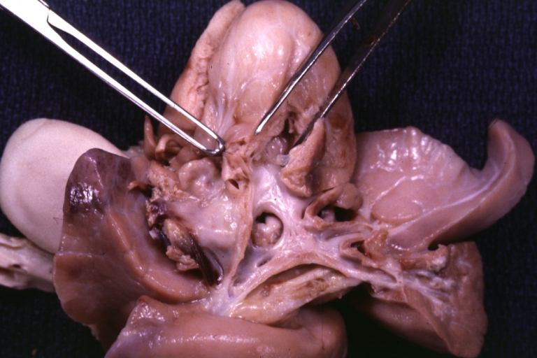
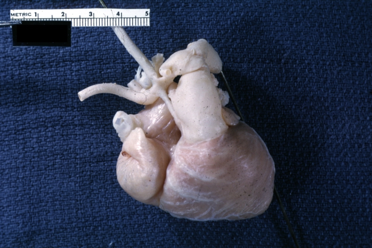
Gross Pathology
-
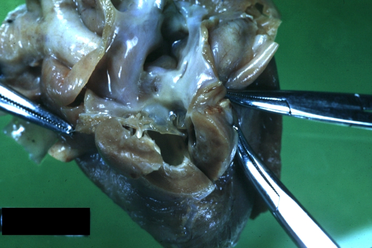 Hypoplastic left ventricle
 -
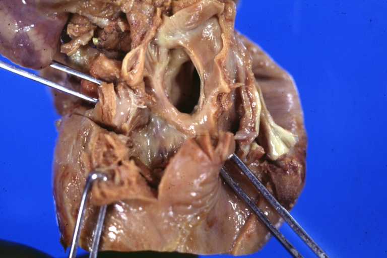 Hypoplastic left ventricle
 -
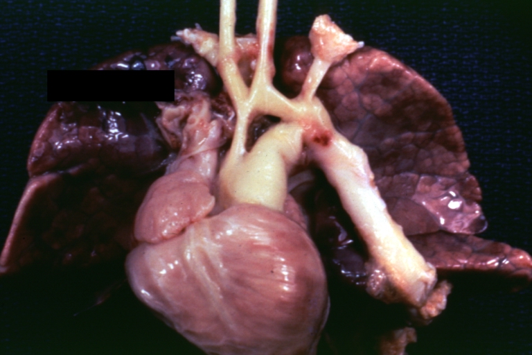 Hypoplastic left ventricle
 -
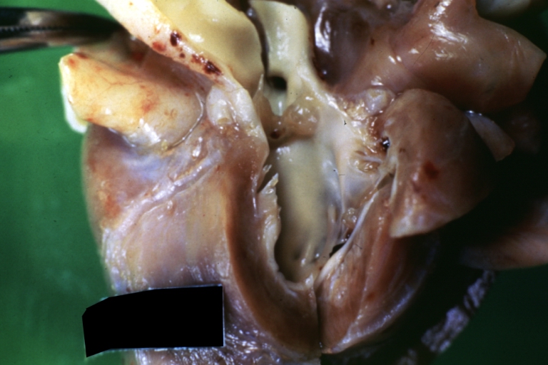 Hypoplastic left ventricle
 -
 Hypoplastic left ventricle
 -
 Hypoplastic left ventricle


Associated Conditions
- HLHS is seen in patients Turner syndrome, Trisomy 18, Trisomy 13 and Jacobsen syndrome
- Anomalous pulmonary venous connection
- Coarctation of the aorta
- Complete atrioventricular canal
- Coronary artery abnormalities (especially in patients with aortic atresia and mitral stenosis)
- Persistent left superior vena cava
- Endocardial fibroelastosis (especially in patients with aortic atresia and mitral stenosis)
Looking for the patient version?
© 2026 MyEClinic – IFTM Institut für Telematik in der Medizin GmbH