
Japanese encephalitis
For patient information, click here.
Editor-In-Chief: C. Michael Gibson, M.S., M.D. [1]; Associate Editor(s)-in-Chief: Anthony Gallo, B.S. [2]
Synonyms and keywords: JE; JEV; Japanese B encephalitis virus; Japanese B encephalitis
Overview
Editor-In-Chief: C. Michael Gibson, M.S., M.D. [1]; Associate Editor(s)-in-Chief: Anthony Gallo, B.S. [2]
Overview
Japanese encephalitis is a moderate infection of the central nervous system. Japanese encephalitis virus is a Group IV positive-sense ssRNA virus within the Flaviviridae family of viruses, and the genus Flavivirus. Japanese encephalitis virus is closely related to Yellow fever virus, Dengue virus, West Nile virus, and St. Louis encephalitis virus.[1] Japanese encephalitis is also known as an arbovirus, or an arthropod-borne virus. Japanese encephalitis virus is usually transmitted via mosquitos to the human host. Japanese encephalitis must be differentiated from other diseases that cause nondescript symptoms, which include fever, headache, and vomiting. Prognosis is generally poor. Approximately 20-30% of patients progress to mortality. Among patients who survive, approximately 50% suffer severe neurological, cognitive, or psychological deficits.[2] The diagnostic method of choice for Japanese encephalitis is laboratory testing. Laboratory findings consistent with the diagnosis of Japanese encephalitis include detection of IgM antibodies in serum and cerebrospinal fluid, moderate leukocytosis, mild anemia, and hyponatremia. There is no treatment for Japanese encephalitis; the mainstay of therapy is supportive care.[3] There are approximately 15 vaccines available for the prevention of Japanese encephalitis.
Historical Perspective
Japanese encephalitis was first discovered in 1871 following a large number of recurring outbreaks in the summer months. In 1930, the first vaccine was developed to prevent Japanese encephalitis; today, there are approximately 15 vaccines available.[4]
Classification
Japanese encephalitis virus is a Group IV positive-sense ssRNA virus within the Flaviviridae family of viruses, and the genus Flavivirus. Japanese encephalitis virus is closely related to Yellow fever virus, Dengue virus, West Nile virus, and St. Louis encephalitis virus.[1] Japanese encephalitis is also known as an arbovirus, or an arthropod-borne virus.
Pathophysiology
Japanese encephalitis virus is usually transmitted via mosquitos to the human host. Japanese encephalitis virus contains positive-sense viral RNA; this RNA has its genome directly utilized as if it were mRNA, producing a single protein which is modified by host and viral proteins to form the various proteins needed for replication. Transmission to humans requires mosquito species capable of creating a “bridge” between infected animals and uninfected humans; this occurs when humans become part of the enzootic cycle. The incubation period is 5-15 days.[5] Humans are dead-end hosts for the virus, meaning there is an insufficient amount of Japanese encephalitis virus in the blood stream to infect a mosquito; there is also no evidence of person to person spread.[6]
Causes
Japanese encephalitis may be caused by Japanese encephalitis virus. Japanese encephalitis virus is closely related to the West Nile virus, Dengue virus, Murray Valley encephalitis virus, and St. Louis encephalitis virus.[1]
Differentiating Japanese encephalitis from Other Diseases
Japanese encephalitis must be differentiated from other diseases that cause nondescript symptoms, which include fever, headache, and vomiting, such as meningitis, malaria, and other demyelinating diseases.[7][8]
Epidemiology and Demographics
The incidence of Japanese encephalitis in patients younger than 19 years old is approximately 10-100 per 100,000 individuals worldwide.[9] The case-fatality rate of Japanese encephalitis is approximately 20-30% worldwide. Japanese encephalitis more commonly affects individuals younger than 18 years of age. Japanese encephalitis usually affects individuals of the Asian race. Japanese encephalitis is most commonly observed in the summer months within temperate climate zones. Japanese encephalitis is a common disease that tends to affect approximately 30,000 to 70,000 individuals in Asia annually.[6]
Risk Factors
The most potent risk factor in the development of Japanese encephalitis is residing in Southeast Asia and Western Pacific regions. Other risk factors include summer season, outdoor recreational activities, and contact with mosquitos, birds, and pigs.[10][11]
Natural History, Complications, and Prognosis
If left untreated, 50% of patients with Japanese encephalitis may progress to develop severe neurological deficits, such as deafness, hemiparesis, and aphasia. Common complications of Japanese encephalitis include seizures, coma, and spastic paralysis. Prognosis is generally poor. Approximately 20-30% of patients progress to mortality. Among patients who survive, approximately 50% suffer severe neurological, cognitive, or psychological deficits.[2]
Diagnosis
History and Symptoms
Most patients infected with Japanese encephalitis remain asymptomatic; 1% of infected individuals develop clinical disease. The incubation period for Japanese encephalitis is usually 5-15 days. Common symptoms of Japanese encephalitis include fever, headache, and vomiting, neck stiffness, stupor, disorientation, coma, tremors, and spastic (but rarely flaccid) paralysis.[12][13]
Physical Examination
Patients with Japanese encephalitis are usually ill-appearing. Physical examination of patients with Japanese encephalitis is usually remarkable for meningism, fever, and convulsions. Signs of Japanese encephalitis which develop during the acute encephalitic stage include neck rigidity, cachexia, hemiparesis, convulsions, and an elevated body temperature between 38-41°C. The classical description of Japanese encephalitis includes a Parkinsonian syndrome with masklike facies, tremor, cogwheel rigidity, and choreoathetoid movements. Acute flaccid paralysis, with clinical and pathological features similar to those of poliomyelitis, has also been associated with Japanese encephalitis.
Laboratory Findings
The diagnostic method of choice for Japanese encephalitis is laboratory testing. Laboratory findings consistent with the diagnosis of Japanese encephalitis include detection of IgM antibodies in serum and cerebrospinal fluid, moderate leukocytosis, mild anemia, and hyponatremia. Cerebrospinal fluid typically has a mild to moderate pleocytosis with a lymphocytic predominance, slightly elevated protein, and normal ratio to plasma glucose. Because humans have low or undetectable levels of viremia by the time distinctive clinical symptoms are recognized, virus isolation and nucleic acid amplification tests are insensitive and should not be used for ruling out a diagnosis of Japanese encephalitis.[14]
CT
On CT scan, Japanese encephalitis is characterized by symmetric or asymmetric bilateral thalamic hypodensities, and subacute or chronic hemorrhagic lesions.
MRI
MRI is the imaging modality of choice for Japanese encephalitis. On MRI, Japanese encephalitis is characterized by symmetric or asymmetric bilateral thalamic hypodensities, and subacute or chronic hemorrhagic lesions.
Other Imaging Studies
On EEG, Japanese encephalitis is characterized by theta and delta coma, burst suppression, epileptiform activity, and occasionally alpha coma.[5]
Treatment
Medical Therapy
There is no treatment for Japanese encephalitis; the mainstay of therapy is supportive care.[3]
Primary Prevention
Effective measures for the primary prevention of Japanese encephalitis include mosquito population control, avoidance of mosquitos, and immunization. Mosquito control has been difficult to achieve in rural settings. Avoidance of exposure is also difficult, as Culex mosquitos are active during day hours. Immunization is the only effective method for sustainable control. Routine immunization of school-age children is currently in use in Korea, Japan, China, Thailand, and Taiwan. The introduction of the Japanese encephalitis vaccine into the Expanded Program of Immunization has helped curb the disease in countries like Thailand, Vietnam, Sri Lanka, and China.[15]
Secondary Prevention
There are no secondary preventive measures available for Japanese encephalitis.
References
- ↑ 1.0 1.1 1.2 Flavivirus. SIB Swiss Institute of Bioinformatics (2015). http://viralzone.expasy.org/viralzone/all_by_species/24.html Accessed on April 12, 2016
- ↑ 2.0 2.1 Khandaker G, Zurynski Y, Buttery J, Marshall H, Richmond PC, Dale RC; et al. (2012). “Neurologic complications of influenza A(H1N1)pdm09: surveillance in 6 pediatric hospitals”. Neurology. 79 (14): 1474–81. doi:10.1212/WNL.0b013e31826d5ea7. PMC 4098823. PMID 22993280.
- ↑ 3.0 3.1 Japanese encephalitis. National Health Service United Kingdom (2016). http://www.nhs.uk/Conditions/Japanese-encephalitis/Pages/Introduction.aspx Accessed on April 12, 2016.
- ↑ Paulke-Korinek M, Kollaritsch H (2008). “Japanese encephalitis and vaccines: past and future prospects”. Wien Klin Wochenschr. 120 (19-20 Suppl 4): 15–9. doi:10.1007/s00508-008-1071-9. PMID 19066766.
- ↑ 5.0 5.1 Japanese Encephalitis Symptoms and Treatment. Centers for Disease Control and Prevention (CDC), National Center for Emerging and Zoonotic Infectious Diseases, Division of Vector-Borne Diseases. (2015) http://www.cdc.gov/japaneseencephalitis/symptoms/ Accessed on April 12, 2016.
- ↑ 6.0 6.1 Japanese encephalitis – Fact sheet No 386. World Health Organization (WHO) (2015) http://www.who.int/mediacentre/factsheets/fs386/en/ Accessed on April 12, 2016
- ↑ M.D. JE, Dolin R, Blaser MJ. Mandell, Douglas, and Bennett’s Principles and Practice of Infectious Diseases, Expert Consult Premium Edition. Saunders; 2014.
- ↑ Kennedy PG (2004). “Viral encephalitis: causes, differential diagnosis, and management”. J Neurol Neurosurg Psychiatry. 75 Suppl 1: i10–5. PMC 1765650. PMID 14978145.
- ↑ Kollaritsch H, Paulke-Korinek M, Dubischar-Kastner K (2009). “IC51 Japanese encephalitis vaccine”. Expert Opin Biol Ther. 9 (7): 921–31. doi:10.1517/14712590903042282. PMID 19527110.
- ↑ Solomon T (2006). “Control of Japanese encephalitis–within our grasp?”. N Engl J Med. 355 (9): 869–71. doi:10.1056/NEJMp058263. PMID 16943399.
- ↑ Japanese encephalitis – Frequently Asked Questions. CDC Centers for Disease Control and Prevention. (2015) http://www.cdc.gov/japaneseencephalitis/qa/index.html Accessed on April 12, 2016
- ↑ Japanese Encephalitis, Clinical and Laboratory Evaluation. Centers for Disease Control and Prevention (2015). http://www.cdc.gov/japaneseencephalitis/healthcareproviders/healthcareproviders-clinlabeval.html Accessed on April 14, 2016.
- ↑ Centers for Disease Control and Prevention (CDC) (2012). “Expanding poliomyelitis and measles surveillance networks to establish surveillance for acute meningitis and encephalitis syndromes–Bangladesh, China, and India, 2006-2008”. MMWR Morb Mortal Wkly Rep. 61 (49): 1008–11. PMID 23235298.
- ↑ Japanese Encephalitis Diagnostic Testing. Centers for Disease Control and Prevention (CDC), National Center for Emerging and Zoonotic Infectious Diseases, Division of Vector-Borne Diseases. (2015) http://www.cdc.gov/japaneseencephalitis/healthcareproviders/healthcareproviders-diagnostic.html Accessed on April 19, 2016.
- ↑ Tauber E, Dewasthaly S (2008). “Japanese encephalitis vaccines–needs, flaws and achievements”. Biol Chem. 389 (5): 547–50. PMID 18953721.
Historical Perspective
Editor-In-Chief: C. Michael Gibson, M.S., M.D. [1]; Associate Editor(s)-in-Chief: Anthony Gallo, B.S. [2]
Overview
Japanese encephalitis was first discovered in 1871 following a large number of recurring outbreaks in the summer months. In 1930, the first vaccine was developed to prevent Japanese encephalitis; today, there are approximately 15 vaccines available.
Historical Perspective
Japanese encephalitis was first discovered in 1871 following a large number of recurring outbreaks in the summer months. There have been several outbreaks of Japanese encephalitis, which occurred in 1927, 1934, and 1935.[1] The association between Culex tritaeniorhynchus as a vector and the involvement of birds and pigs as reservoir hosts for Japanese encephalitis was made in 1938.[2] In 1930, the first vaccine was developed to prevent Japanese encephalitis; today, there are approximately 15 vaccines available.[3]
References
- ↑ Erlanger TE, Weiss S, Keiser J, Utzinger J, Wiedenmayer K (2009). “Past, present, and future of Japanese encephalitis”. Emerg Infect Dis. 15 (1): 1–7. doi:10.3201/eid1501.080311. PMC 2660690. PMID 19116041.
- ↑ Tsai TF, Yu, XY. 1994 Japanese encephalitis vaccines. Plotkin SA, Mortimer EA, eds. Vaccines. Second edition. Philadel- pha: W. B. Saunders, 671–714.
- ↑ Paulke-Korinek M, Kollaritsch H (2008). “Japanese encephalitis and vaccines: past and future prospects”. Wien Klin Wochenschr. 120 (19-20 Suppl 4): 15–9. doi:10.1007/s00508-008-1071-9. PMID 19066766.
Classification
Editor-In-Chief: C. Michael Gibson, M.S., M.D. [1]; Associate Editor(s)-in-Chief: Anthony Gallo, B.S. [2]
Overview
Japanese encephalitis may be classified according to location of the disease into 2 subtypes: systemic or encephalitic. Japanese encephalitis may also be classified according to neuroinvasiveness of the disease into 2 subtypes: neuroinvasive and non-neuroinvasive. Japanese encephalitis is also known as an arbovirus, or an arthropod-borne virus.
Classification
Japanese encephalitis may be classified according to location of the disease into 2 subtypes: systemic or encephalitic. Japanese encephalitis may also be classified according to neuroinvasiveness of the disease into 2 subtypes: neuroinvasive and non-neuroinvasive. Japanese encephalitis virus is a Group IV positive-sense ssRNA virus within the Flaviviridae family of viruses, and the genus Flavivirus. Japanese encephalitis virus is closely related to Yellow Fever virus, Dengue virus, West Nile virus, and St. Louis encephalitis virus.[1] Japanese encephalitis is also known as an arbovirus, or an arthropod-borne virus.
References
- ↑ Flavivirus. SIB Swiss Institute of Bioinformatics (2015). http://viralzone.expasy.org/viralzone/all_by_species/24.html Accessed on April 12, 2016
Pathophysiology
Editor-In-Chief: C. Michael Gibson, M.S., M.D. [1]; Associate Editor(s)-in-Chief: Anthony Gallo, B.S. [2]
Overview
Japanese encephalitis virus is usually transmitted via mosquitos to the human host. Japanese encephalitis virus contains positive-sense viral RNA; this RNA has its genome directly utilized as if it were mRNA, producing a single protein which is modified by host and viral proteins to form the various proteins needed for replication. Transmission to humans requires mosquito species capable of creating a “bridge” between infected animals and uninfected humans; this occurs when humans become part of the enzootic cycle. The incubation period is 5-15 days. Humans are dead-end hosts for the virus, meaning there is an insufficient amount of Japanese encephalitis virus in the blood stream to infect a mosquito; there is also no evidence of person to person spread.
Pathophysiology
Japanese encephalitis virus is usually transmitted via mosquitos to the human host. Japanese encephalitis virus contains positive-sense viral RNA; this RNA has its genome directly utilized as if it were mRNA, producing a single protein which is modified by host and viral proteins to form the various proteins needed for replication. The following table is a summary of the Japanese encephalitis virus:[1][2]
| Characteristic | Data |
|---|---|
| Nucleic acid | RNA |
| Sense | ssRNA(+) |
| Virion | Enveloped |
| Capsid | Spherical |
| Symmetry | Yes; T=3-like organization; icosahedral-like |
| Capsid monomers | Unknown |
| Envelope length (diameter) | 50 nm |
| Additional envelope information | Mature virons contain 2 virus-encoded membrane proteins (M and E); immature virons contain a protein precursor |
| Genome shape | Linear |
| Genome length | 10-11 kb |
| Nucleotide cap | Yes |
| Polyadenylated tail | No; a loop structure is formed instead |
| Incubation period | 5-15 days |
Japanese encephalitis is contracted by the bite of an infected mosquito, primarily Culex tritaeniorhynchus. Japanese encephalitis virus circulates between a mosquito vector and either pigs or water birds (such as herons) in Southeast Asia.[3] Transmission to humans requires mosquito species capable of creating a “bridge” between infected animals and uninfected humans; this occurs when humans become part of the enzootic cycle. The incubation period is 5-15 days.[4] Humans are dead-end hosts for the virus, meaning there is an insufficient amount of Japanese encephalitis virus in the blood stream to infect a mosquito; there is also no evidence of person to person spread.[5]
Japanese encephalitis virus is transmitted in the following pattern:[1]
- Attachment of the viral envelope protein E to host receptors mediates internalization into the host cell by clathrin-mediated endocytosis, or by apoptotic mimicry.
- Fusion of virus membrane with host endosomal membrane. RNA genome is released into the cytoplasm.
- The positive-sense ssRNA virus is translated into a polyprotein, which is cleaved into all structural and non-structural proteins necessary for RNA synthesis (replication and transcription).
- Replication takes place at the surface of endoplasmic reticulum in cytoplasmic viral factories. A dsRNA genome is synthesized from the genomic ssRNA(+).
- The dsRNA genome is transcribed/replicated, thereby providing viral mRNAs/new ssRNA(+) genomes.
- Virus assembly occurs at the endoplasmic reticulum. The virion buds via the host endosomal sorting complexes required for transport (ESCRT), and is sent to the Golgi apparatus.
- The prM protein is cleaved in the Golgi, thereby maturing the virion which is fusion competent.
- New virions are released by exocytosis.
On microscopic histopathological analysis, the enveloped, spherical, and icosahedral-like virion shape are characteristic findings of Japanese encephalitis.
Gallery
-
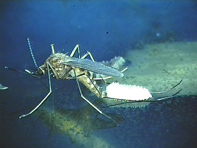 Culex mosquito laying eggs. (Photograph by Richard G. Weber)
 -
![Diagram illustrates the methods by which the arbovirus, Japanese encephalitis virus (JEV) reproduces and amplifies itself in the avian populations, and is subsequently transmitted to human beings as the dead end host. From Public Health Image Library (PHIL). [6]](https://www.wikidoc.org/images/6/6a/Flavivirus02.jpeg) Diagram illustrates the methods by which the arbovirus, Japanese encephalitis virus (JEV) reproduces and amplifies itself in the avian populations, and is subsequently transmitted to human beings as the dead end host. From Public Health Image Library (PHIL). [6]
Diagram illustrates the methods by which the arbovirus, Japanese encephalitis virus (JEV) reproduces and amplifies itself in the avian populations, and is subsequently transmitted to human beings as the dead end host. From Public Health Image Library (PHIL). [6]

![Diagram illustrates the methods by which the arbovirus, Japanese encephalitis virus (JEV) reproduces and amplifies itself in the avian populations, and is subsequently transmitted to human beings as the dead end host. From Public Health Image Library (PHIL). [6]](https://www.wikidoc.org/index.php/File%3AFlavivirus02.jpeg)
References
- ↑ 1.0 1.1 Flavivirus. SIB Swiss Institute of Bioinformatics. (2015) http://viralzone.expasy.org/viralzone/all_by_species/24.html Accessed on April 12, 2016
- ↑ Japanese encephalitis – Frequently Asked Questions. CDC Centers for Disease Control and Prevention. (2015) http://www.cdc.gov/japaneseencephalitis/qa/index.html Accessed on April 12, 2016
- ↑ Japanese encephalitis. National Health Service United Kingdom (2016). http://www.nhs.uk/Conditions/Japanese-encephalitis/Pages/Introduction.aspx Accessed on April 12, 2016.
- ↑ Japanese Encephalitis Symptoms and Treatment. Centers for Disease Control and Prevention (CDC), National Center for Emerging and Zoonotic Infectious Diseases, Division of Vector-Borne Diseases. (2015) http://www.cdc.gov/japaneseencephalitis/symptoms/ Accessed on April 12, 2016.
- ↑ Japanese encephalitis – Fact sheet No 386. World Health Organization (WHO) (2015) http://www.who.int/mediacentre/factsheets/fs386/en/ Accessed on April 12, 2016
- ↑ “Public Health Image Library (PHIL)”.
Causes
Editor-In-Chief: C. Michael Gibson, M.S., M.D. [1]; Associate Editor(s)-in-Chief: Anthony Gallo, B.S. [2]
Overview
Japanese encephalitis may be caused by Japanese encephalitis virus. Japanese encephalitis virus is closely related to the West Nile virus, Dengue virus, Murray Valley encephalitis virus, and St. Louis encephalitis virus.[1]
Causes
Japanese encephalitis may be caused by Japanese encephalitis virus. Japanese encephalitis virus is closely related to the West Nile virus, Dengue virus, Murray Valley encephalitis virus, and St. Louis encephalitis virus.[1]
References
- ↑ 1.0 1.1 Flavivirus. SIB Swiss Institute of Bioinformatics (2015). http://viralzone.expasy.org/viralzone/all_by_species/24.html Accessed on April 12, 2016
Differentiating Japanese encephalitis from other Diseases
Editor-In-Chief: C. Michael Gibson, M.S., M.D. [1]; Associate Editor(s)-in-Chief: Anthony Gallo, B.S. [2]
Overview
Japanese encephalitis must be differentiated from other diseases that cause nondescript symptoms, which include fever, headache, and vomiting, such as meningitis, brain abscess, and other demyelinating diseases.
Differentiating Japanese encephalitis from Other Diseases
Japanese encephalitis must be differentiated from other diseases that cause fever, headache, and vomiting, such as:[1][2][3][4][5]
| Disease | Similarities | Differentials |
|---|---|---|
| Meningitis | Classic triad of fever, nuchal rigidity, and altered mental status | Photophobia, phonophobia, rash associated with meningococcemia, concomitant sinusitis or otitis, swelling of the fontanelle in infants (0-6 months) |
| Brain abscess | Fever, headache, hemiparesis | Varies depending on the location of the abscess; clinically, visual disturbance including papilledema, decreased sensation; on imaging, a lesion demonstrates both ring enhancement and central restricted diffusion |
| Demyelinating diseases | Ataxia, lethargy | Multiple sclerosis: clinically, nystagmus, internuclear ophthalmoplegia, Lhermitte’s sign; on imaging, well-demarcated ovoid lesions with possible T1 hypointensities (“black holes”)
Acute disseminated encephalomyelitis: clinically, somnolence, myoclonic movements, and hemiparesis; on imaging, diffuse or multi-lesion enhancement, with indistinct lesion borders |
| Substance abuse | Tremor, headache, altered mental status | Varies depending on type of substance: prior history, drug-seeking behavior, attention-seeking behavior, paranoia, sudden panic, anxiety, hallucinations |
| Electrolyte disturbance | Fatigue, headache, nausea | Varies depending on deficient ions; clinically, edema, constipation, hallucinations; on EKG, abnormalities in T wave, P wave, QRS complex; possible presentations include arrhythmia, dehydration, renal failure |
| Stroke | Ataxia, aphasia, dizziness | Varies depending on classification of stroke; presents with positional vertigo, high blood pressure, extremity weakness |
| Intracranial hemorrhage | Headache, coma, dizziness | Lobar hemorrhage, numbness, tingling, hypertension, hemorrhagic diathesis |
| Trauma | Headache, altered mental status | Amnesia, loss of consciousness, dizziness, concussion, contusion |
References
- ↑ M.D. JE, Dolin R, Blaser MJ. Mandell, Douglas, and Bennett’s Principles and Practice of Infectious Diseases, Expert Consult Premium Edition. Saunders; 2014.
- ↑ Kennedy PG (2004). “Viral encephalitis: causes, differential diagnosis, and management”. J Neurol Neurosurg Psychiatry. 75 Suppl 1: i10–5. PMC 1765650. PMID 14978145.
- ↑ Arboviral Infections (arthropod-borne encephalitis, eastern equine encephalitis, St. Louis encephalitis, California encephalitis, Powassan encephalitis, West Nile encephalitis). New York State Department of Health (2006). https://www.health.ny.gov/diseases/communicable/arboviral/fact_sheet.htm Accessed on February 23, 2016
- ↑ Eckstein C, Saidha S, Levy M (2012). “A differential diagnosis of central nervous system demyelination: beyond multiple sclerosis”. J Neurol. 259 (5): 801–16. doi:10.1007/s00415-011-6240-5. PMID 21932127.
- ↑ De Kruijk JR, Twijnstra A, Leffers P (2001). “Diagnostic criteria and differential diagnosis of mild traumatic brain injury”. Brain Inj. 15 (2): 99–106. doi:10.1080/026990501458335. PMID 11260760.
Epidemiology and Demographics
Editor-In-Chief: C. Michael Gibson, M.S., M.D. [1]; Associate Editor(s)-in-Chief: Anthony Gallo, B.S. [2]
Overview
The incidence of Japanese encephalitis in patients younger than 19 years old is approximately 10-100 per 100,000 individuals worldwide. The case-fatality rate of Japanese encephalitis is approximately 20-30% worldwide. Japanese encephalitis more commonly affects individuals younger than 18 years of age. Japanese encephalitis usually affects individuals of the Asian race. Japanese encephalitis is most commonly observed in the summer months within temperate climate zones. Japanese encephalitis is a common disease that tends to affect approximately 30,000 to 70,000 individuals in Asia annually.
Epidemiology and Demographics
Prevalence
There are 24 countries in Southeast Asia and Western Pacific regions that have Japanese encephalitis virus transmission risk, a population of over 3 billion people.[1]
Incidence
The incidence of Japanese encephalitis in patients younger than 19 years old is approximately 10-100 per 100,000 individuals worldwide.[2]
Case-Fatality Rate
The case-fatality rate of Japanese encephalitis is approximately 20-30% worldwide.[3]
Age
Japanese encephalitis more commonly affects individuals younger than 18 years of age. In rural areas where Japanese encephalitis is endemic, seroprevalance approaches 100%; approximately 1 in 300 infected children develop Japanese encephalitis.[4]
Race
Japanese encephalitis usually affects individuals of the Asian race.
Seasonal
Japanese encephalitis is most commonly observed in the summer months within temperate climate zones. Year-round transmission is observed in tropical climate zones.[5]
Geographical Location
The majority of Japanese encephalitis cases are reported in South and Southeast Asia. There have been suspected cases on the Indian subcontinent.
Developed Countries
Japanese encephalitis is a rare disease that tends to sporadically cause outbreaks which affect U.S. territories in the Western Pacific.
Developing Countries
Japanese encephalitis is a common disease that tends to affect approximately 30,000 to 70,000 individuals in Asia annually.[1]

References
- ↑ 1.0 1.1 Japanese encephalitis – Fact sheet No 386. World Health Organization (WHO) (2015) http://www.who.int/mediacentre/factsheets/fs386/en/ Accessed on April 12, 2016
- ↑ Kollaritsch H, Paulke-Korinek M, Dubischar-Kastner K (2009). “IC51 Japanese encephalitis vaccine”. Expert Opin Biol Ther. 9 (7): 921–31. doi:10.1517/14712590903042282. PMID 19527110.
- ↑ Japanese encephalitis – Frequently Asked Questions. CDC Centers for Disease Control and Prevention. (2015) http://www.cdc.gov/japaneseencephalitis/qa/index.html Accessed on April 12, 2016
- ↑ Centers for Disease Control and Prevention (CDC) (2012). “Expanding poliomyelitis and measles surveillance networks to establish surveillance for acute meningitis and encephalitis syndromes–Bangladesh, China, and India, 2006-2008”. MMWR Morb Mortal Wkly Rep. 61 (49): 1008–11. PMID 23235298.
- ↑ Vaughn DW, Hoke CH (1992). “The epidemiology of Japanese encephalitis: prospects for prevention”. Epidemiol Rev. 14: 197–221. PMID 1337744.
Risk Factors
Editor-In-Chief: C. Michael Gibson, M.S., M.D. [1]; Associate Editor(s)-in-Chief: Anthony Gallo, B.S. [2]
Overview
The most potent risk factor in the development of Japanese encephalitis is residing in Southeast Asia and Western Pacific regions. Other risk factors include summer season, outdoor recreational activities, and contact with mosquitos, birds, and pigs.
Risk Factors
Common risk factors in the development of Japanese encephalitis are:[1][2][3]
- Residents or military in Southeast Asia and Western Pacific regions
- Summer season
- Outdoor recreational activities
- Accommodations in endemic ares that lack air conditioning, bed nets, or window screens
- Age extremes
- Contact with:
- Mosquitos
- Birds
- Pigs
References
- ↑ Solomon T (2006). “Control of Japanese encephalitis–within our grasp?”. N Engl J Med. 355 (9): 869–71. doi:10.1056/NEJMp058263. PMID 16943399.
- ↑ Japanese encephalitis – Frequently Asked Questions. CDC Centers for Disease Control and Prevention. (2015) http://www.cdc.gov/japaneseencephalitis/qa/index.html Accessed on April 12, 2016
- ↑ Japanese encephalitis – Fact sheet No 386. World Health Organization (WHO) (2015) http://www.who.int/mediacentre/factsheets/fs386/en/ Accessed on April 12, 2016
Natural History, Complications and Prognosis
Editor-In-Chief: C. Michael Gibson, M.S., M.D. [1]; Associate Editor(s)-in-Chief: Anthony Gallo, B.S. [2]
Overview
If left untreated, 50% of patients with Japanese encephalitis may progress to develop severe neurological deficits, such as deafness, hemiparesis, and aphasia. Common complications of Japanese encephalitis include seizures, coma, and spastic paralysis. Prognosis is generally poor. Approximately 20-30% of patients progress to mortality. Among patients who survive, approximately 50% suffer severe neurological, cognitive, or psychological deficits.
Natural History
If left untreated, 50% of patients with Japanese encephalitis may progress to develop severe neurological deficits, such as deafness, hemiparesis, and aphasia.[1][2]
Complications
Common complications of Japanese encephalitis include:[3][4]
Prognosis
Prognosis is generally poor. Approximately 20-30% of patients progress to mortality. Among patients who survive, approximately 50% suffer severe neurological, cognitive, or psychological deficits.[1][3]
References
- ↑ 1.0 1.1 Khandaker G, Zurynski Y, Buttery J, Marshall H, Richmond PC, Dale RC; et al. (2012). “Neurologic complications of influenza A(H1N1)pdm09: surveillance in 6 pediatric hospitals”. Neurology. 79 (14): 1474–81. doi:10.1212/WNL.0b013e31826d5ea7. PMC 4098823. PMID 22993280.
- ↑ M.D. JE, Dolin R, Blaser MJ. Mandell, Douglas, and Bennett’s Principles and Practice of Infectious Diseases, Expert Consult Premium Edition. Saunders; 2014.
- ↑ 3.0 3.1 Japanese encephalitis – Fact sheet No 386. World Health Organization (WHO) (2015) http://www.who.int/mediacentre/factsheets/fs386/en/ Accessed on April 12, 2016
- ↑ Japanese Encephalitis, Clinical and Laboratory Evaluation. Centers for Disease Control and Prevention (2015). http://www.cdc.gov/japaneseencephalitis/healthcareproviders/healthcareproviders-clinlabeval.html Accessed on April 14, 2016.
Diagnosis
Diagnosis
History and Symptoms | Physical Examination | Laboratory Findings | CT | MRI | Other Diagnostic Studies
Treatment
Treatment
Medical Therapy | Primary Prevention | Secondary Prevention | Cost-Effectiveness of Therapy | Future or Investigational Therapies
External Links
External Links
Acknowledgements
Acknowledgements
The content on this page was first contributed by: C. Michael Gibson, M.S., M.D.
Looking for the patient version?
© 2026 MyEClinic – IFTM Institut für Telematik in der Medizin GmbH