
Mesothelioma chest x ray
Editor-In-Chief: C. Michael Gibson, M.S., M.D. [1]Associate Editor(s)-in-Chief: Parminder Dhingra, M.D. [2], Sujit Routray, M.D. [3]
Overview
Overview
Chest x-rays are non-specific in a case of mesothelioma, demonstrating a pleural opacity which may extend around and encase the lung.[1]
Chest X Ray
Chest X Ray
- Chest x-rays are non-specific in a case of mesothelioma, may demonstrate a opacity in the pleural lining which may extend around and encase the lung.[1]
- X-ray may show evidence of mediastinum shift resulting from the reduced volume of the affected hemithorax.
- Rib destruction or extension beyond the lateral and anterior margins of the chest wall may be evident.
- Mediastinal lymph node enlargement and pleural effusion may also be seen.[1]
Gallery
Gallery
-
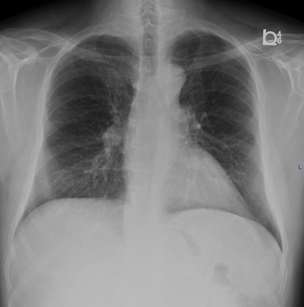 CXR of a patient came for evaluation for left sided pleural effusion with possible asbestos exposure demonstrates very subtle left sided pleural thickening.<ref name=cxrimagemesothelioma1>Image courtesy of Dr. A.Prof Frank Gaillard. Radiopaedia (original file here). Creative Commons BY-SA-NC

References
References
- ↑ 1.0 1.1 1.2 Mesothelioma. Dr Bruno Di Muzio and A.Prof Frank Gaillard et al. Radiopaedia 2016. http://radiopaedia.org/articles/mesothelioma. Accessed on 13th January, 2016
Looking for the patient version?
© 2026 MyEClinic – IFTM Institut für Telematik in der Medizin GmbH