
Mesothelioma
For patient information click here
Editor-In-Chief: C. Michael Gibson, M.S., M.D. [1]Associate Editor(s)-in-Chief: Feham Tariq, MD [2]Parminder Dhingra, M.D. [3], Sujit Routray, M.D. [4]
Synonyms and keywords: Mesotheliomas; Benign mesothelioma; Malignant mesothelioma; Peritoneal mesothelioma; Pericardial mesothelioma; Pleural mesothelioma; Malignant pleural mesothelioma; Mesothelioma of the pleura
Overview
Editor-In-Chief: C. Michael Gibson, M.S., M.D. [1]Associate Editor(s)-in-Chief: Sujit Routray, M.D. [2]
Overview
Mesothelioma is a rare, highly aggressive cancer which arises from the mesothelial cells which form the lining of the pleural, and less frequently the peritoneal, pericardial, and tunica vaginalis cavities. Mesothelioma is a form of cancer that is most commonly caused by exposure to asbestos. Wagner et al. were the first to discover the association between asbestos exposure and development of mesothelioma. Mesothelioma may be classified into several subtypes based on the location (pleural, peritoneal, pericardial, cystic/multicystic, and tunica vaginalis testis), histology (epithelial, sarcomatoid, and biphasic), and potential to spread (benign and malignant). Asbestos causes DNA damage directly by mechanically interfering with the segregation of chromosomes during mitosis and indirectly by inducing mesothelial cells and macrophages, to release mutagenic reactive oxygen and nitrogen species. Asbestos fibres have been shown to alter the function and secretory properties of macrophages, ultimately creating conditions which favor the development of mesothelioma. Following asbestos phagocytosis, macrophages generate increased amounts of hydroxyl radicals, which are normal by-products of cellular anaerobic metabolism. However, these free radicals are also known clastogenic and membrane-active agents thought to promote asbestos carcinogenicity. These oxidants can participate in the oncogenic process by directly and indirectly interacting with DNA, modifying membrane-associated cellular events, including oncogene activation and perturbation of cellular antioxidant defences. Genes involved in the pathogenesis of mesothelioma include BAP1, CDKN2A, WT1, NF2, and TP53. On gross pathology, pleural mesothelioma is characterized by discrete plaques and nodules that coalesce to produce a sheet-like tumor, with the pleural surface seeding of malignant mesothelioma cells. Based on the histology, mesothelioma may be classified into 3 subtypes: epithelial, sarcomatoid, and biphagic. Mesothelioma is demonstrated by positivity to tumor markers, such as calretinin, epithelial membrane antigen, cytokeratin, and mesothelin. Mesothelioma must be differentiated from pleural effusion, lung cancer, pulmonary tuberculosis, peritoneal tuberculosis, pseudomyxoma peritonei, constrictive pericarditis, ovarian cystadenoma, and mesothelial hyperplasia of the testis. If left untreated, mesothelioma may progress to develop dyspnea, dysphagia, pleural effusion, thrombophlebitis, constrictive pericarditis, recurrent hydrocele, and metastases, depending on the site involved. Complications of mesothelioma include pleural effusion, spinal cord compression, Horner’s syndrome, superior vena cava syndrome, hyperviscocity syndrome, pericardial effusion, cardiac tamponade, heart failure, ascites, and recurrent hydrocele. The prognosis of mesothelioma depends on the cell subtype. According to the Union for International Cancer Control staging system, there are four stages of pleural mesothelioma based on the primary tumor, lymph nodes, and metastasis: stage I (IA, IB), stage II, stage III, and stage IV. There is no established staging system for peritoneal mesothelioma. When evaluating a patient for mesothelioma, you should take a detailed history of the presenting symptom (onset, duration, and progression), other associated symptoms, and a thorough family and past medical history review. Other specific areas of focus include a history of any exposure to asbestos or radiation. Symptoms of mesothelioma may not appear until 20 to 50 years after exposure to asbestos. Symptoms of mesothelioma include chest pain, shortness of breath, cough, abdominal pain, palpitation, hemoptysis, and swelling in the legs. CT is the most commonly used modality for the assessment of mesothelioma and is able to stage the disease accurately in majority of the patients. Chemotherapy is one of the mainstay of therapy for mesothelioma. Other therapies for mesothelioma include radiotherapy, surgery, and supportive care. Effective measures for the primary prevention of mesothelioma include removal of asbestos from schools and other public buildings. Secondary prevention strategies following mesothelioma include regular checkups, chest x-rays, and/or CT scans in high-risk individuals.
Historical Perspective
Wagner et al. were the first to discover the association between asbestos exposure and development of mesothelioma.
Classification
Mesothelioma may be classified into several subtypes based on the location (pleural, peritoneal, pericardial, cystic/multicystic, and tunica vaginalis testis), histology (epithelial, sarcomatoid, and biphasic), and potential to spread (benign and malignant).
Pathophysiology
Asbestos causes DNA damage directly by mechanically interfering with the segregation of chromosomes during mitosis and indirectly by inducing mesothelial cells and macrophages, to release mutagenic reactive oxygen and nitrogen species. Asbestos fibres have been shown to alter the function and secretory properties of macrophages, ultimately creating conditions which favor the development of mesothelioma. Following asbestos phagocytosis, macrophages generate increased amounts of hydroxyl radicals, which are normal by-products of cellular anaerobic metabolism. However, these free radicals are also known clastogenic and membrane-active agents thought to promote asbestos carcinogenicity. These oxidants can participate in the oncogenic process by directly and indirectly interacting with DNA, modifying membrane-associated cellular events, including oncogene activation and perturbation of cellular antioxidant defences. Genes involved in the pathogenesis of mesothelioma include BAP1, CDKN2A, WT1, NF2, and TP53. On gross pathology, pleural mesothelioma is characterized by discrete plaques and nodules that coalesce to produce a sheet-like tumor, with the pleural surface seeding of malignant mesothelioma cells. Based on the histology, mesothelioma may be classified into 3 subtypes: epithelial, sarcomatoid, and biphagic. Mesothelioma is demonstrated by positivity to tumor markers, such as calretinin, epithelial membrane antigen, cytokeratin, and mesothelin.
Causes
Common causes of mesothelioma include asbestos-fibre exposure, erionite-fibre exposure, Simian virus 40, and radiation exposure.
Differentiating Mesothelioma from other Diseases
Mesothelioma must be differentiated from pleural effusion, lung cancer, pulmonary tuberculosis, peritoneal tuberculosis, pseudomyxoma peritonei, constrictive pericarditis, ovarian cystadenoma, and mesothelial hyperplasia of the testis.
Epidemiology and Demographics
Mesothelioma is a rare disease which accounts for 5-28% of all malignancies that involve the pleura. The incidence of mesothelioma is estimated to be 3,000 cases annually. The incidence of pleural mesothelioma is approximately 1 per 100,000 individuals in the United States. Males are more commonly affected with mesothelioma than females. The male to female ratio is approximately 3 to 1. The incidence of mesothelioma increases with age; the median age at diagnosis for pleural mesothelioma and peritoneal mesothelioma are 74 years and 68 years, respectively. There is no racial predilection to mesothelioma.
Risk Factors
The most potent risk factor in the development of mesothelioma is asbestos exopsure. Other risk factors include erionite-fibre exposure, Simian virus 40, and radiation exposure.
Screening
There is insufficient evidence to recommend routine screening for mesothelioma.
Natural History, Complications and Prognosis
If left untreated, mesothelioma may progress to develop dyspnea, dysphagia, pleural effusion, thrombophlebitis, constrictive pericarditis, recurrent hydrocele, and metastases, depending on the site involved. Complications of mesothelioma include pleural effusion, spinal cord compression, Horner’s syndrome, superior vena cava syndrome, hyperviscocity syndrome, pericardial effusion, cardiac tamponade, heart failure, ascites, and recurrent hydrocele. The prognosis of mesothelioma depends on the cell subtype.
Diagnosis
Staging
According to the Union for International Cancer Control staging system, there are four stages of pleural mesothelioma based on the primary tumor, lymph nodes, and metastasis: stage I (IA, IB), stage II, stage III, and stage IV. There is no established staging system for peritoneal mesothelioma.
History and Symptoms
When evaluating a patient for mesothelioma, you should take a detailed history of the presenting symptom (onset, duration, and progression), other associated symptoms, and a thorough family and past medical history review. Other specific areas of focus include a history of any exposure to asbestos or radiation. Symptoms of mesothelioma may not appear until 20 to 50 years after exposure to asbestos. Symptoms of mesothelioma include chest pain, shortness of breath, cough, abdominal pain, palpitation, hemoptysis, and swelling in the legs.
Physical Examination
Common physical examination findings of mesothelioma include dullness on percussion and decreased breath sounds on ausculatation of the lung, ascites, abdominal mass, hemoptysis, pedal edema, and murmurs.
Laboratory Findings
Laboratory findings consistent with the diagnosis of mesothelioma include abnormal pleural fluid analysis (decreased pleural pH and pleural fluid/serum glucose ratio).
Chest X Ray
Chest x-rays are of limited utility and are non-specific in a case of mesothelioma, demonstrating a pleural opacity which may extend around and encase the lung.
CT
Chest CT scan may be diagnostic of mesothelioma. CT is the most commonly used modality for the assessment of mesothelioma and is able to stage the disease accurately in majority of the patients.
MRI
MRI, although not routinely used, may have a role in refining the staging and better delineating the extent of the disease in surgical candidates especially with regard to chest wall and diaphragmatic invasion. Findings on MRI include iso- to slightly hyperintense on T1-weighted images and iso- to hyperintense on T2-weighted images. There is enhancement on contrast administration.
Other Diagnostic Studies
Other diagnostic studies for mesothelioma include laparoscopy, thoracoscopy, pleuroscopy, biopsy, position emission tomography scan, and fluorescence in situ hybridization.
Treatment
Medical Therapy
Chemotherapy is one of the mainstay of therapy for mesothelioma. Other therapies for mesothelioma include radiotherapy, surgery, and supportive care.
Surgery
The feasibility of surgery depends on the stage of mesothelioma at diagnosis.
Summary of Treatment for Pleural Mesothelioma
The treatment options vary for the different stages, subtypes, and resectability of pleural mesothelioma. The types of treatment given are also based on the unique needs of the individual with cancer.
Summary of Treatment for Peritoneal Mesothelioma
The types of treatment given are based on the unique needs of the individual with cancer. Peritoneal mesothelioma is a locally aggressive disease that is difficult to treat. The goal of the treatment is to control the disease for as long as possible, manage symptoms, and improve the person’s quality of life.
Primary Prevention
Effective measures for the primary prevention of mesothelioma include removal of asbestos from schools and other public buildings.
Secondary Prevention
Secondary prevention strategies following mesothelioma include regular checkups, chest x-rays, and/or CT scans in high-risk individuals.
References
Historical Perspective
Editor-In-Chief: C. Michael Gibson, M.S., M.D. [1]Associate Editor(s)-in-Chief: Parminder Dhingra, M.D. [2], Sujit Routray, M.D. [3], Fatima Shaukat, MD [4]
Overview
The association between mesothelioma and asbestos exposure was first described by Dr. Wagner in his seminal study of South African miners. It was discovered later that people who live near to factors and mines developed mesothelioma despite not working in the mines.
Historical Perspective
- The association between mesothelioma and exposure to asbestos fibers was elucidated by Dr. Wagner in his seminal study of South African miners.[1]
- In the town of Wittenoom, asbestos-containing mine waste was used to cover schoolyards and playgrounds.
- In 1965, an article in the British Journal of Industrial Medicine established that people who lived in the neighborhoods of asbestos factories and mines, but did not work in them, had contracted mesothelioma.
- Despite proof that the dust associated with asbestos mining and milling causes asbestos related disease, mining began at Wittenoom in 1943 and continued until 1966. In 1974 the first public warnings of the dangers of blue asbestos were published in a cover story called “Is this Killer in Your Home?” in Australia’s Bulletin magazine. In 1978, the Western Australian Government decided to phase out the town of Wittenoom, following the publication of a Health Dept. booklet, “The Health Hazard at Wittenoom”, containing the results of air sampling and an appraisal of worldwide medical information.
- By 1979 the first writs for negligence related to Wittenoom were issued against CSR and its subsidiary ABA, and the Asbestos Diseases Society was formed to represent the Wittenoom victims.
- In the landmark study by Vogelzang et al., cisplatin and pemetrexed led to an objective response rate of 41% and median survival of 12.1 months, compared to 17% and 9.3 months in patients treated with cisplatin alone.[2]
Legal issues
- The first lawsuits against asbestos manufacturers were in 1929.
- Since then, many lawsuits have been filed against asbestos manufacturers and employers, for neglecting to implement safety measures after the links between asbestos, asbestosis, and mesothelioma became known (some reports seem to place this as early as 1898).
- The liability resulting from the sheer number of lawsuits and people affected has reached billions of dollars.
- The amounts and method of allocating compensation have been the source of many court cases, and government attempts at resolution of existing and future cases.
References
- ↑ Thomas, Anish; Chen, Yuanbin; Yu, Tinghui; Gill, Ammara; Prasad, Vinay (2015). “Distinctive clinical characteristics of malignant mesothelioma in young patients”. Oncotarget. 6 (18): 16766–16773. doi:10.18632/oncotarget.4414. ISSN 1949-2553.
- ↑ Saint-Pierre, Mathieu D.; Pease, Christopher; Mithoowani, Hamid; Zhang, Tinghua; Nicholas, Garth A.; Laurie, Scott A.; Wheatley-Price, Paul (2015). “Malignant Pleural Mesothelioma Outcomes in the Era of Combined Platinum and Folate Antimetabolite Chemotherapy”. Lung Cancer International. 2015: 1–7. doi:10.1155/2015/590148. ISSN 2090-3197.
Classification
Editor-In-Chief: C. Michael Gibson, M.S., M.D. [1]Associate Editor(s)-in-Chief: Parminder Dhingra, M.D. [2], Sujit Routray, M.D. [3]
Overview
Mesothelioma may be classified into several subtypes based on the location (pleural, peritoneal, pericardial, cystic/multicystic, and tunica vaginalis testis), histology (epithelial, sarcomatoid, and biphasic), and potential to spread (benign and malignant).
Classification
Based on the location, mesothelioma may be classified into 5 subtypes:[1]
- Pleural mesothelioma
- Peritoneal mesothelioma
- Pericardial mesothelioma
- Cystic/multicystic mesothelioma
- Tunica vaginalis testis mesothelioma
Based on the histology, mesothelioma may be classified into 3 subtypes:[1]
- Epithelial
- Sarcomatoid
- Biphasic (mixed)
Based on the potential to spread, mesothelioma may be classified into 2 subtypes:[2]
- Malignant mesothelioma
- Benign mesothelioma
- Fibrous tumor of the pleura: Fibrous tumor of the pleura is a benign tumor that forms in the visceral or parietal pleura. This type of tumor can occur in both sexes. A benign fibrous tumor of the pleura can cause symptoms similar to malignant mesothelioma, such as shortness of breath. It may also advance to form a malignant tumor. A benign fibrous tumor of the pleura can recur after surgical resection.
- Multicystic mesothelioma: Multicystic mesothelioma forms several benign cysts in the peritoneum. This type of mesothelioma most often occurs in femlaes. It may also be called benign cystic mesothelioma.
- Adenomatoid mesothelioma: Adenomatoid mesothelioma is a benign tumor that usually develops in the mesothelium of the male and female genital system. In males, this benign tumor often starts in the epididymis. In femlaes, adenomatoid mesothelioma starts in the Fallopian tubes.[3]
References
- ↑ 1.0 1.1 Mesothelioma. Dr Bruno Di Muzio and A.Prof Frank Gaillard et al. Radiopaedia 2016. http://radiopaedia.org/articles/mesothelioma. Accessed on February 8, 2016
- ↑ What is mesothelioma. Canadian cancer society 2016. http://www.cancer.ca/en/cancer-information/cancer-type/mesothelioma/mesothelioma/?region=on. Accessed on February 8, 2016
- ↑ Benign tumours of the mesothelium. Canadian cancer society 2016. http://www.cancer.ca/en/cancer-information/cancer-type/mesothelioma/mesothelioma/benign-tumours/?region=on. Accessed on February 8, 2016
Pathophysiology
Editor-In-Chief: C. Michael Gibson, M.S., M.D. [1]; Associate Editor(s)-in-Chief: Feham Tariq, MD [2],Sujit Routray, M.D. [3]
Overview
Asbestos causes DNA damage directly by mechanically interfering with the segregation of chromosomes during mitosis and indirectly by inducing mesothelial cells and macrophages, to release mutagenic reactive oxygen and nitrogen species. Asbestos fibres have been shown to alter the function and secretory properties of macrophages, ultimately creating conditions which favor the development of mesothelioma. Following asbestos phagocytosis, macrophages generate increased amounts of hydroxyl radicals, which are normal by-products of cellular anaerobic metabolism. However, these free radicals are also known clastogenic and membrane-active agents thought to promote asbestos carcinogenicity. These oxidants can participate in the oncogenic process by directly and indirectly interacting with DNA, modifying membrane-associated cellular events, including oncogene activation and perturbation of cellular antioxidant defences. Genes involved in the pathogenesis of mesothelioma include BAP1, CDKN2A, WT1, NF2, and TP53. On gross pathology, pleural mesothelioma is characterized by discrete plaques and nodules that coalesce to produce a sheet-like tumor, with the pleural surface seeding of malignant mesothelioma cells. Based on the histology, mesothelioma may be classified into 3 subtypes: epithelial, sarcomatoid, and biphagic. Mesothelioma is demonstrated by positivity to tumor markers, such as calretinin, epithelial membrane antigen, cytokeratin, and mesothelin.
Pathogenesis
The pathogenesis of mesothelioma is influenced by the following factors:[1][2][3][4]
Types of Asbestos fibres
There are two types of asbestos fibers:
- Amphibole (sharp, rod-like)
- Serpentine
Role of Asbestos fibers
Asbestos fibers play the following role in the development of mesothelioma:
- The most common type of fibres found in US are the serpentine fibers, which are considered less carcinogenic than the amphibole ones.
- Following inhalation from the environment, these fibres are trapped in the lower third zone of the lung.
- They are phagocytosed by the mesothelial cells and initiate an oncogenic cascade of events which include:
- Asbestos causes DNA damage directly by mechanically interfering with the segregation of chromosomes during mitosis and indirectly by inducing mesothelial cells and macrophages, to release mutagenic reactive oxygen and nitrogen species.
- Asbestos fibres have been shown to alter the function and secretory properties of macrophages, ultimately creating conditions which favor the development of mesothelioma.
- Following asbestos phagocytosis, macrophages generate increased amounts of hydroxyl radicals, which are normal by-products of cellular anaerobic metabolism.
- However, these free radicals are also known clastogenic and membrane-active agents thought to promote asbestos carcinogenicity. These oxidants can participate in the oncogenic process by directly and indirectly interacting with DNA, modifying membrane-associated cellular events, including oncogene activation and perturbation of cellular antioxidant defences.
- The mesothelium consists of a single layer of flattened to cuboidal cells forming the epithelial lining of the serous cavities of the body including the peritoneal, pericardial and pleural cavities.
- Deposition of asbestos fibres in the parenchyma of the lung may result in the penetration of the visceral pleura from where the fibre can then be carried to the pleural surface, thus leading to the development of malignant mesothelial plaques.
- The processes leading to the development of peritoneal mesothelioma remain unresolved, although it has been proposed that asbestos fibres from the lung are transported to the abdomen and associated organs via the lymphatic system.
- Additionally, asbestos fibres may be deposited in the gut after ingestion of sputum contaminated with asbestos fibres.
- Asbestos also may possess immunosuppressive properties. For example, chrysotile fibres have been shown to depress the in-vitro proliferation of phytohemagglutinin-stimulated peripheral blood lymphocytes, suppress natural killer cell lysis, and significantly reduce lymphokine-activated killer cell viability and recovery.
- Furthermore, genetic alterations in asbestos-activated macrophages may result in the release of potent mesothelial cell mitogens such as platelet-derived growth factor (PDGF) and transforming growth factor-β (TGF-β) which in turn, may induce the chronic stimulation and proliferation of mesothelial cells after injury by asbestos fibres.
Radiation therapy
Survivors of radiation therapy have been found to develop mesothelioma according to several studies. Following cancers have been studied post-radiation therapy which are seen to develop mesothelioma:[5][6][7]
- Non-Hodgkins lymphoma[8]
- Testicular cancer
- Breast cancer
Smoking
- Smoking has a synergistic effect on asbestos fibre inhalation in the pathogenesis of mesothelioma.
Genetics
- Development of mesothelioma is the result of multiple genetic mutations.
- Genes involved in the pathogenesis of mesothelioma include:[9][10][11][12][13][14]
- Asbestos has also been shown to mediate the entry of foreign DNA into target cells. Incorporation of this foreign DNA may lead to mutations and oncogenesis, by several possible mechanisms:
- Inactivation of tumor suppressor genes
- Activation of oncogenes
- Activation of proto-oncogenes due to incorporation of foreign DNA containing a promoter region
- Activation of DNA repair enzymes, which may be prone to error
- Activation of telomerase
- Prevention of apoptosis
Gross Pathology
The following features are seen on gross pathology of mesothelioma:[15]
- On gross pathology, pleural mesothelioma is characterized by discrete plaques and nodules that coalesce to produce a sheet-like tumor, with the pleural surface seeding of malignant mesothelioma cells.
- The growth usually starts at the inferior margins of the pleura and may invade the diaphragm.
- The lung and interlobar fissures may also be involved.
Gallery
-
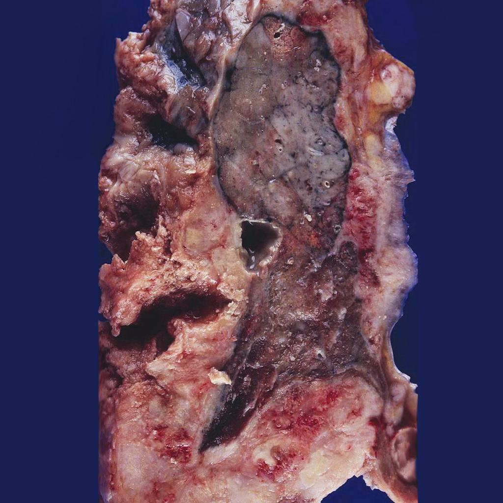 Mesothelioma completely encasing the lung.<ref name=grosspictureofmesotheliomaimage1>Image courtesy of Dr. Yale Rosen. Radiopaedia (original file here). Creative Commons BY-SA-NC

Microscopic Pathology
Based on the histology, mesothelioma may be classified into 3 subtypes:[16]
- Epithelial
- Sarcomatoid
- Biphasic (mixed)
Immunohistochemistry
- The cytological and histological diagnosis can be difficult, with mesothelial hyperplasia and metastatic adenocarcinoma appearing similar. Specific markers may be helpful in the diagnosis of mesothelioma.[17]
- Mesothelioma is demonstrated by positivity to tumor markers, such as:[17][18]
- Calretinin
- Epithelial membrane antigen
- Cytokeratin
- Mesothelin
- WT1
- Podoplanin
- Osteopontin
- HBME-1
References
- ↑ Selikoff IJ, Hammond EC, Seidman H (1980). “Latency of asbestos disease among insulation workers in the United States and Canada”. Cancer. 46 (12): 2736–40. PMID 7448712.
- ↑ Heintz NH, Janssen-Heininger YM, Mossman BT (2010). “Asbestos, lung cancers, and mesotheliomas: from molecular approaches to targeting tumor survival pathways”. Am J Respir Cell Mol Biol. 42 (2): 133–9. doi:10.1165/rcmb.2009-0206TR. PMC 2822975. PMID 20068227.
- ↑ Thomas, Anish; Chen, Yuanbin; Yu, Tinghui; Gill, Ammara; Prasad, Vinay (2015). “Distinctive clinical characteristics of malignant mesothelioma in young patients”. Oncotarget. 6 (18): 16766–16773. doi:10.18632/oncotarget.4414. ISSN 1949-2553.
- ↑ Shukla A, Gulumian M, Hei TK, Kamp D, Rahman Q, Mossman BT (2003). “Multiple roles of oxidants in the pathogenesis of asbestos-induced diseases”. Free Radic Biol Med. 34 (9): 1117–29. PMID 12706492.
- ↑ Travis LB, Fosså SD, Schonfeld SJ, McMaster ML, Lynch CF, Storm H; et al. (2005). “Second cancers among 40,576 testicular cancer patients: focus on long-term survivors”. J Natl Cancer Inst. 97 (18): 1354–65. doi:10.1093/jnci/dji278. PMID 16174857.
- ↑ Matesich SM, Shapiro CL (2003). “Second cancers after breast cancer treatment”. Semin Oncol. 30 (6): 740–8. PMID 14663775.
- ↑ Tward JD, Wendland MM, Shrieve DC, Szabo A, Gaffney DK (2006). “The risk of secondary malignancies over 30 years after the treatment of non-Hodgkin lymphoma”. Cancer. 107 (1): 108–15. doi:10.1002/cncr.21971. PMID 16708354.
- ↑ Travis LB, Curtis RE, Glimelius B, Holowaty E, Van Leeuwen FE, Lynch CF; et al. (1993). “Second cancers among long-term survivors of non-Hodgkin’s lymphoma”. J Natl Cancer Inst. 85 (23): 1932–7. PMID 8230284.
- ↑ Ladanyi M (2005). “Implications of P16/CDKN2A deletion in pleural mesotheliomas”. Lung Cancer. 49 Suppl 1: S95–8. doi:10.1016/j.lungcan.2005.03.017. PMID 15950811.
- ↑ Kumar-Singh S, Segers K, Rodeck U, Backhovens H, Bogers J, Weyler J; et al. (1997). “WT1 mutation in malignant mesothelioma and WT1 immunoreactivity in relation to p53 and growth factor receptor expression, cell-type transition, and prognosis”. J Pathol. 181 (1): 67–74. doi:10.1002/(SICI)1096-9896(199701)181:1<67::AID-PATH723>3.0.CO;2-Z. PMID 9072005.
- ↑ Ladanyi M, Zauderer MG, Krug LM, Ito T, McMillan R, Bott M; et al. (2012). “New strategies in pleural mesothelioma: BAP1 and NF2 as novel targets for therapeutic development and risk assessment”. Clin Cancer Res. 18 (17): 4485–90. doi:10.1158/1078-0432.CCR-11-2375. PMC 3432735. PMID 22825583.
- ↑ Andujar P, Pairon JC, Renier A, Descatha A, Hysi I, Abd-Alsamad I; et al. (2013). “Differential mutation profiles and similar intronic TP53 polymorphisms in asbestos-related lung cancer and pleural mesothelioma”. Mutagenesis. 28 (3): 323–31. doi:10.1093/mutage/get008. PMID 23435014.
- ↑ Opitz I, Soltermann A, Abaecherli M, Hinterberger M, Probst-Hensch N, Stahel R; et al. (2008). “PTEN expression is a strong predictor of survival in mesothelioma patients”. Eur J Cardiothorac Surg. 33 (3): 502–6. doi:10.1016/j.ejcts.2007.09.045. PMID 18248818.
- ↑ Murakami H, Mizuno T, Taniguchi T, Fujii M, Ishiguro F, Fukui T; et al. (2011). “LATS2 is a tumor suppressor gene of malignant mesothelioma”. Cancer Res. 71 (3): 873–83. doi:10.1158/0008-5472.CAN-10-2164. PMID 21245096.
- ↑ Mesothelioma. CGMH.ORG 2016. https://www1.cgmh.org.tw/intr/intr5/c6700/OBGYN/f/web/Mesothelioma/index.htm. Accessed on February 15, 2016
- ↑ Mesothelioma. Dr Bruno Di Muzio and A.Prof Frank Gaillard et al. Radiopaedia 2016. http://radiopaedia.org/articles/mesothelioma. Accessed on February 8, 2016
- ↑ 17.0 17.1 Pathology of mesothelioma. Dr Bruno Di Muzio and A.Prof Frank Gaillard et al. Radiopaedia 2016. http://radiopaedia.org/articles/mesothelioma. Accessed on February 10, 2016
- ↑ Immunohistochemistry of mesothelioma. Wikipedia 2016. https://en.wikipedia.org/wiki/Mesothelioma. Accessed on February 12, 2016
Causes
Editor-In-Chief: C. Michael Gibson, M.S., M.D. [1]Associate Editor(s)-in-Chief: Fatima Shaukat, MD [2]Parminder Dhingra, M.D. [3]
Overview
Common causes of mesothelioma include asbestos-fibre exposure, erionite-fibre exposure, Simian virus 40, radiation exposure and genetic predesposition[1][2]
Causes
Mesothelioma is caused by:[1][2]
- Asbestos-fibre exposure: causes majority of cases
- Erionite-fibre exposure
- Simian virus 40 (SV40)
- Radiation exposure
- Genetic predisposition
Asbestos
- The strongest and most common cause for mesothelioma is occupational exposure to asbestos, which has been widely used in building materials and many industries.[1]
- Asbestos is naturally occurring group of mineral consisting of very fine,long and thin fibers.
- Being so fine, they can be inhaled easily and may be lodged in the smallest airways of the lung and the mesothelium, eventually leading to pleural mesothelioma.
- Sometimes, instead of inhalation, the fibers are coughed up and swallowed. This way they can settle in the peritoneum to cause peritoneal mesothelioma.
- According to the International Agency for Research on Cancer (IARC) and the US National Toxicology Program, all forms of asbestos are known to cause cancer. Most people with mesothelioma have a history of asbestos exposure.
- Mesothelioma has a long latency period, which means it usually doesn’t develop for 15–40, or more, years after exposure to asbestos.
- There are 2 types of exposure to asbestos:
- Direct exposure affects people who come into contact with asbestos directly. This may include:[1] [3]
- Asbestos mines/mills workers
- Asbestos products/asbestos-based products producers
- Construction workers, carpenters and painters
- Shipyard workers
- Cement manufacturers
- Insulation workers
- Electricians and heating tradespeople
- Plumbers
- Demolition workers
- Automotive industry workers, including brake and clutch repair workers
- Indirect exposure affects people who come in contact with asbestos in other ways. This may include:[1]
- Family members who are exposed to asbestos from fibres brought home on a worker’s clothing
- People who live in or near an asbestos factory or mine
- Direct exposure affects people who come into contact with asbestos directly. This may include:[1] [3]
- The risk of developing mesothelioma is related to how much asbestos a person was exposed to and how long the exposure lasted.
- People exposed at an early age, for a long period of time and to greater amounts of asbestos are more likely to develop mesothelioma.
- Occasionally, mesothelioma develops in people who have never been exposed to asbestos.[4]
Erionite
- Erionite is another naturally occurring mineral, happens to be a known human carcinogen associated with development of pleural and peritoneal mesothelioma.
- The group of minerals, erionite belongs to is called zeolites.[1]
- Zeolites are chemically related to asbestos and erionite has asbestos-like fibres.
- Erionite is common in the soil in parts of Turkey and materials made with erionite are used in construction in these regions.
- High rates of mesothelioma in these areas are due to exposure to this mineral.[1]
Ionizing Radiation
- People who have been treated with radiation therapy to the chest or abdomen for lymphoma, breast cancer, lung cancer, or other cancers can cause mesothelioma.[1]
- Although the risk of mesothelioma is higher in people who have been treated with radiation therapy, mesothelioma is likely to occur in only a very small number of these people.
- There have been reports linking mesothelioma to Thorotrast (thorium dioxide). Thorotrast is a contrast medium once used for imaging tests, but it is no longer used.[1]
Simian virus 40
- Infection with SV40 may cause mesothelioma.
- Evidence suggested the contamination of polio vaccines with SV40 from 1955 to 1963, which may have contributed to some cases of mesothelioma.
- It is thought that SV40 may act as a co-factor with asbestos in causing mesothelioma.[1]
Genetic predesposition
- Based on the fact that malignant mesothelioma clustering was observed in few families, a study was conducted in 2012 on caucasian american population to determine the genetic association.
- It was found that people with a somatic germline mutation in their BAP1 gene is associated with higher risk of developing mesothelioma and uveal melanoma.[2]
References
- ↑ 1.00 1.01 1.02 1.03 1.04 1.05 1.06 1.07 1.08 1.09 Risk factors for mesothelioma. Canadian cancer society 2016. http://www.cancer.ca/en/cancer-information/cancer-type/mesothelioma/risks/?region=on. Accessed on February 8, 2016
- ↑ 2.0 2.1 2.2 Testa JR, Cheung M, Pei J, Below JE, Tan Y, Sementino E; et al. (2011). “Germline BAP1 mutations predispose to malignant mesothelioma”. Nat Genet. 43 (10): 1022–5. doi:10.1038/ng.912. PMC 3184199. PMID 21874000.
- ↑ Gennaro V, Finkelstein MM, Ceppi M, Fontana V, Montanaro F, Perrotta A; et al. (2000). “Mesothelioma and lung tumors attributable to asbestos among petroleum workers”. Am J Ind Med. 37 (3): 275–82. PMID 10642417.
- ↑ Henderson DW, Rödelsperger K, Woitowitz HJ, Leigh J (2004). “After Helsinki: a multidisciplinary review of the relationship between asbestos exposure and lung cancer, with emphasis on studies published during 1997-2004”. Pathology. 36 (6): 517–50. PMID 15841689.
Differentiating Mesothelioma from other Diseases
Editor-In-Chief: C. Michael Gibson, M.S., M.D. [1]Associate Editor(s)-in-Chief: Parminder Dhingra, M.D. [2], Sujit Routray, M.D. [3]
Overview
Mesothelioma must be differentiated from pleural effusion, lung cancer, pulmonary tuberculosis, peritoneal tuberculosis, pseudomyxoma peritonei, constrictive pericarditis, ovarian cystadenoma, and mesothelial hyperplasia of the testis.
Differentiating Mesothelioma from other Diseases
Differentiating Pleural Mesothelioma from other Diseases
Pleural mesothelioma must be differentiated from:[1][2][1][2][3][4][5][6][7]
- Pleural effusion
- Benign asbestos-related pleural disease
- Peripheral bronchogenic carcinoma
- Pleural fibrosis from infective/inflammatory source (e.g. actinomycetes, tuberculosis)
- Primary pleural tumors
- Secondary lesions that can involve the pleura
- Pleural metastases
- Thymoma with pleural invasion
- Pericardial tumors with pleural invasion
- Ewing sarcoma of chest wall with pleural invasion
Differentiating Peritoneal Mesothelioma from other Diseases
Peritoneal mesothelioma must be differentiated from:[3][4]
- Peritoneal carcinomatosis
- Pseudomyxoma peritonei
- Lymphoma with peritoneal involvement
- Peritoneal involvement with tuberculosis
- Mesenteric panniculitis
- Primary peritoneal adenomatoid tumor
- Primary peritoneal papillary serous carcinoma
- Primary peritoneal serous borderline tumor
- Diffuse peritoneal leiomyomatosis
- Desmoplastic small round cell tumor arising from the peritoneum
- Solitary fibrous tumor arising the peritoneum
- Peritoneal lymphangioma
- Peritoneal inclusion cyst
- Spontaneous bacterial peritonitis (SBP)
Differentiating Pericardial Mesothelioma from other Diseases
Pericardial mesothelioma must be differentiated from:[5]
- Heart failure
- Coronary heart disease
- Constrictive pericarditis
- Cardiomyopathy
- Tuberculosis pericarditis
- Cardiac tamponade
- Intra-atrial myxoma
Differentiating Multicystic Mesothelioma from other Diseases
Multicystic mesothelioma must be differentiated from:[6]
- Abdominopelvic cystic lymphangioma
- Ovarian cystadenoma
- Ovarian cystadenocarcinoma
- Endometriosis
- Cystic teratoma
- Cystic mucinous tumor of pancreas
Differentiating Tunica Vaginalis Testis Mesothelioma from other Diseases
Tunica vaginalis testis mesothelioma must be differentiated from:[7]
- Mesothelial hyperplasia of the testis
- Adenomatoid tumor of the testis
- Rete testis adenocarcinoma
- Serous papillary tumors of the testis and epididymis
- Pleomorphic rhabdomyosarcoma
- Malignant fibrous histiocytoma
- Germ cell tumors of the testis
Differentiating peritoneal Mesothelioma from other Diseases
| Disease | Prominent clinical findings | Lab tests | Tratment | |
|---|---|---|---|---|
| Primary peritonitis | Spontaneous bacterial peritonitis |
|
|
|
| Tuberculous peritonitis |
|
|
| |
| Continuous Ambulatory Peritoneal Dialysis (CAPD peritonitis) |
|
|
| |
| Secondary peritonitis | Acute bacterial secondary peritonitis |
|
| |
| Biliary peritonitis |
|
|||
| Tertiary peritonitis |
|
|
| |
| Familial Mediterranean fever (periodic peritonitis, familial paroxysmal polyserositis) |
|
| ||
| Granulomatous peritonitis |
|
|
| |
| Sclerosing encapsulating peritonitis |
|
|||
| Intraperitoneal abscesses |
|
|
| |
| Peritoneal mesothelioma |
|
|
| |
| peritoneal carcinomatosis |
|
|||
References
- ↑ 1.0 1.1 Differential diagnosis of mesothelioma. Dr Bruno Di Muzio and A.Prof Frank Gaillard et al. Radiopaedia 2016. http://radiopaedia.org/articles/mesothelioma. Accessed on February 12, 2016
- ↑ 2.0 2.1 Dr Yuranga Weerakkody et al. Radiopaedia 2016. http://radiopaedia.org/articles/pleural-tumours. Accessed on February 12, 2016
- ↑ 3.0 3.1 Differential diagnosis of peritoneal mesothelioma. Dr Alexandra Stanislavsky et al. Radiopaedia 2016. http://radiopaedia.org/articles/peritoneal-mesothelioma. Accessed on February 12, 2016
- ↑ 4.0 4.1 Primary peritoneal neoplasms. Dr Praveen Jha and Radswiki et al. Radiopaedia 2016. http://radiopaedia.org/articles/primary-peritoneal-neoplasms. Accessed on February 12, 2016
- ↑ 5.0 5.1 Seek a Second Opinion to Avoid Misdiagnosis of Pericardial Mesothelioma. Asbestos.com 2016. http://www.asbestos.com/mesothelioma/pericardial.php. Accessed on February 12, 2016
- ↑ 6.0 6.1 Differential diagnosis of multicystic mesothelioma. Dr Aditya Shetty and Dr Yuranga Weerakkody et al. Raiopaedia 2016. http://radiopaedia.org/articles/multicystic-mesothelioma. Accessed on February 12, 2016
- ↑ 7.0 7.1 Chekol, Seble S; Sun, Chen-Chin (2012). “Malignant Mesothelioma of the Tunica Vaginalis Testis: Diagnostic Studies and Differential Diagnosis”. Archives of Pathology & Laboratory Medicine. 136 (1): 113–117. doi:10.5858/arpa.2010-0550-RS. ISSN 0003-9985.
Epidemiology and Demographics
Editor-In-Chief: C. Michael Gibson, M.S., M.D. [1]Associate Editor(s)-in-Chief: Fatima Shaukat, MD [2]Parminder Dhingra, M.D. [3], Sujit Routray, M.D. [4]
Overview
Mesothelioma is a rare disease which accounts for 5-28% of all malignancies that involve the pleura. The incidence of mesothelioma is estimated to be 3,000 cases annually. The incidence of pleural mesothelioma is approximately 1 per 100,000 individuals in the United States. Males are more commonly affected with mesothelioma than females. The male to female ratio is approximately 3 to 1. The incidence of mesothelioma increases with age; the median age at diagnosis for pleural mesothelioma and peritoneal mesothelioma are 74 years and 68 years, respectively. There is no racial predilection to mesothelioma.
Epidemiology and Demographics
Prevalence
- Mesothelioma is a rare disease which accounts for 5-28% of all malignancies that involve the pleura.[1]
Incidence
- The incidence of mesothelioma is estimated to be 3,000 cases annually.[2]
- The incidence of pleural mesothelioma is approximately 1 per 100,000 individuals in the United States.[3]
- The incidence of pleural mesothelioma is approximately 1.2 per 100,000 individuals in Canada.[3]
- The incidence of mesothelioma has decreased over several decades in the United States coincident with diminishing occupational asbestos exposure and has remained stable since 2003.[4]
Age
- Males are more commonly affected with mesothelioma than females. The male to female ratio is approximately 3 to 1.[1]
- The incidence of mesothelioma increases with age; the median age at diagnosis for pleural mesothelioma and peritoneal mesothelioma are 74 years and 68 years, respectively.[4]
Race
- There is no racial predilection to mesothelioma.[1]
References
- ↑ 1.0 1.1 1.2 Epidemiology of mesothelioma. Dr Bruno Di Muzio and A.Prof Frank Gaillard et al. Radiopaedia 2016. http://radiopaedia.org/articles/mesothelioma. Accessed on February 8, 2016
- ↑ Philip A. Rascoe, Xiaobo X. Cao and W. Roy Smythe (2012). Molecular Pathogenesis of Malignant Pleural Mesothelioma, Mesotheliomas – Synonyms and Definition, Epidemiology, Etiology, Pathogenesis, Cyto-Histopathological Features, Clinic, Diagnosis, Treatment, Prognosis, Dr Alexander Zubritsky (Ed.), ISBN: 978-953-307-845-8, InTech, Available from: http://www.intechopen.com/books/mesotheliomas-synonyms-and-definition-epidemiology-etiology-pathogenesis-cyto-histopathological-features-clinic-diagnosis-treatment-prognosis/molecular-pathogenesis-of-malignant-pleural-mesothelioma
- ↑ 3.0 3.1 Saint-Pierre, Mathieu D.; Pease, Christopher; Mithoowani, Hamid; Zhang, Tinghua; Nicholas, Garth A.; Laurie, Scott A.; Wheatley-Price, Paul (2015). “Malignant Pleural Mesothelioma Outcomes in the Era of Combined Platinum and Folate Antimetabolite Chemotherapy”. Lung Cancer International. 2015: 1–7. doi:10.1155/2015/590148. ISSN 2090-3197.
- ↑ 4.0 4.1 Thomas, Anish; Chen, Yuanbin; Yu, Tinghui; Gill, Ammara; Prasad, Vinay (2015). “Distinctive clinical characteristics of malignant mesothelioma in young patients”. Oncotarget. 6 (18): 16766–16773. doi:10.18632/oncotarget.4414. ISSN 1949-2553.
Risk Factors
Editor-In-Chief: C. Michael Gibson, M.S., M.D. [1]Associate Editor(s)-in-Chief: Parminder Dhingra, M.D. [2], Sujit Routray, M.D. [3]
Overview
The most potent risk factor in the development of mesothelioma is asbestos exopsure. Other risk factors include erionite-fibre exposure, Simian virus 40, and radiation exposure.[1]
Risk Factors
- The most potent risk factor in the development of mesothelioma is asbestos exopsure.[1]
- Other risk factors include erionite-fibre exposure, Simian virus 40, and radiation exposure.[1]
- Specific risk factors for multicystic mesothelioma include:[2]
References
- ↑ 1.0 1.1 1.2 Mesothelioma. Dr Bruno Di Muzio and A.Prof Frank Gaillard et al.Radiopaedia 2016. http://radiopaedia.org/articles/mesothelioma. Accessed on 13th January, 2016
- ↑ Risk factors for multicystic mesothelioma. Dr Aditya Shetty and Dr Yuranga Weerakkody et al. Radiopaedia 2016. http://radiopaedia.org/articles/multicystic-mesothelioma. Accessed on February 13, 2016
Screening
Editor-In-Chief: C. Michael Gibson, M.S., M.D. [1]Associate Editor(s)-in-Chief: Fatima Shaukat, MD [2]Parminder Dhingra, M.D. [3]
Overview
There is insufficient evidence to recommend routine screening for mesothelioma.
Screening
- There is no universally agreed protocol for screening people who have been exposed to asbestos.
- However some research indicates that the serum osteopontin level might be useful in screening asbestos-exposed people for mesothelioma.
- The level of soluble mesothelin-related protein is elevated in the serum of about 75% of patients at diagnosis and it has been suggested that it may be useful for screening.[1]
References
- ↑ “Soluble mesothelin-related protein–a blood test for mesothelioma” by B. W. Robinson, J. Creaney, R. Lake, A. Nowak, A. W. Musk, N. de Klerk, P. Winzell, K. E. Hellstrom and I. Hellstrom in Lung Cancer (2005) volume 49, pages S109-S111 Template:Entrez Pubmed.
Natural History, Complications and Prognosis
Editor-In-Chief: C. Michael Gibson, M.S., M.D. [1]Associate Editor(s)-in-Chief: Parminder Dhingra, M.D. [2], Sujit Routray, M.D. [3]
Overview
If left untreated, mesothelioma may progress to develop dyspnea, dysphagia, pleural effusion, thrombophlebitis, constrictive pericarditis, recurrent hydrocele, and metastases, depending on the site involved. Complications of mesothelioma include pleural effusion, spinal cord compression, Horner’s syndrome, superior vena cava syndrome, hyperviscocity syndrome, pericardial effusion, cardiac tamponade, heart failure, ascites, and recurrent hydrocele. The prognosis of mesothelioma depends on the cell subtype.[1]
Natural History
- Malignant mesothelioma is usually an aggressive disease.[1]
- The exposure to the asbestos that cause mesothelioma occurrs 25-40 years to appear.
- Metastasis outside the thoracic wall occurs late in the course of pleural mesothelioma. Common sites of metastasis for pleural mesothelioma include:[2]
- Mediastinum
- Pericardium
- Lymph nodes
- Thoracic wall
- Contralateral lung
- Diaphragm
- Peritoneum (the membrane that lines the walls of the abdomen and pelvis, and covers and supports most of the abdominal organs)
- Liver
- Adrenal gland
- Kidney
- Brain
- Peritoneal mesothelioma does not usually spread to the lymph nodes or distant organs. Common sites of metastasis for peritoneal mesothelioma include:[2]
- Serosa of the small and large intestine – can cause a bowel obstruction
- Unilateral or bilateral pleural cavities
- Liver
- Spleen
Complications
Complications of Pleural Mesothelioma
Common complications of pleural mesothelioma include:[3][4][5][6]
- Difficulty swallowing
- Pain caused by pressure on the nerves and spinal cord
- Pleural effusion
- Atelectasis
- Hypercoagulability syndrome
- Spinal cord compression
- Horner’s syndrome
- Superior vena cava syndrome
Complications of Peritoneal Mesothelioma
Common complications of peritoneal mesothelioma include:[7]
- Anorexia
- Weight loss
- Hypercoagulability syndrome
- Ascites
Complications of Pericardial Mesothelioma
Common complications of pericardial mesothelioma include:[8]
Complications of Tunica Vaginalis Testis Mesothelioma
Common complications of tunica vaginalis testis mesothelioma include:[9]
Prognosis
- Prognosis in mesothelioma is difficult to assess consistently because there is great variability in the time before diagnosis and the rate of disease progression.[10]
- The prognostic factors for mesothelioma include:[11]
- Cell subtype: Epithelioid mesothelioma is the most common subtype of mesothelioma and has a better prognosis than sarcomatoid or mixed (biphasic) types. Sarcomatoid subtype has the least favorable prognosis.
- Location of mesothelioma: Pericardial mesothelioma is usually associated with a poorer prognosis than the other types of mesothelioma.
- Surgical removal: Mesothelioma that can be resected has a more favorable prognosis than mesothelioma that has spread too far and is unresectable. Clear surgical margins improve prognosis. Mesothelioma is often a diffuse disease and doesn’t stay localized. This makes it difficult to get clear surgical margins.
- Stage: Stage plays a role in prognosis for people who have surgery. The stage of mesothelioma does not impact survival for people who do not have surgery. Generally, early stage mesothelioma has a better prognosis than more advanced stages. If mesothelioma has spread to the lymph nodes, it is usually associated with a less favorable prognosis.
- Symptoms: The presence of chest pain with pleural mesothelioma or excessive weight loss is associated with a poorer prognosis. Chest pain suggests there may advanced disease that is unresectable.
- Thrombocytosis: Thrombocytosis is associated with a poorer prognosis.
- Leukocytosis: Leukocytosis is associated with a poorer prognosis.
- Performance status: People with a good performance status have a better prognosis than those with a poor performance status.
- Age: Younger people have a better prognosis than older people.
- Sex: Females seem to have a better prognosis than men.
- Lactate dehydrogenase (LDH) level: People with increased LDH blood levels tend to have a less favorable prognosis than those with normal LDH levels. LDH is an enzyme in the blood that can be increased when there is damage to certain tissue or cancer.
- Mesothelioma occurring in germline BAP1 mutation carriers have been reported to be less aggressive clinically and associated with prolonged survival compared with sporadic mesothelioma.[12]
- As discussed earlier, the prognosis of mesothelioma depends on the cell subtype. The median survival time of various subtypes of mesothelioma are tabulated below.[1]
| Location of mesothelioma | Approximate median survival |
|---|---|
| Pleural | 4-18 months |
| Peritoneal | 5-12 months |
| Pericardial | 6 months |
| Tunica vaginalis testis | 23 months |
References
- ↑ 1.0 1.1 1.2 Survival statistics for mesothelioma. Canadian cancer society 2016. http://www.cancer.ca/en/cancer-information/cancer-type/mesothelioma/prognosis-and-survival/survival-statistics/?region=on. Accessed on February 10, 2016
- ↑ 2.0 2.1 If mesothelioma spreads. Canadian cancer society 2016. http://www.cancer.ca/en/cancer-information/cancer-type/mesothelioma/if-cancer-spreads/?region=on. Accessed on February 15, 2016
- ↑ Complications of mesothelioma. Mayo clinic 2016. http://www.mayoclinic.org/diseases-conditions/mesothelioma/basics/complications/con-20026157. Accessed on February 13, 2016
- ↑ Mensi C, Termine L, Garberi A, Meroni S, Levi D, Balzarini L; et al. (2012). “Spinal cord compression: an unusual presentation of malignant pleural mesothelioma. A case report and review of the literature”. Tumori. 98 (4): e92–7. doi:10.1700/1146.12651. PMID 23052177.
- ↑ Minami T, Matsumoto K, Aizawa H, Nakano H, Sugio K, Nakashima Y; et al. (1999). “[Horner’s syndrome in a patient with diffuse malignant pleural mesothelioma]”. Nihon Kokyuki Gakkai Zasshi. 37 (4): 287–90. PMID 10390966.
- ↑ Ragalie GF, Varkey B, Choi H (1983). “Malignant pleural mesothelioma presenting as superior vena cava syndrome”. Can Med Assoc J. 128 (6): 689–91, 740. PMC 1875200. PMID 6825037.
- ↑ Clinical presentation of peritoneal mesothelioma. Dr Alexandra Stanislavsky et al. Radiopaedia 2016. http://radiopaedia.org/articles/peritoneal-mesothelioma. Accessed on February 13, 2016
- ↑ Complications of pericardial mesothelioma. Dr Henry Knipe and Dr Yuranga Weerakkody et al. Radiopaedia 2016. http://radiopaedia.org/articles/pericardial-mesothelioma. Accessed on February 13, 2016
- ↑ Clinical presentation of tunica vaginalis testis mesothelioma. Dr Matt A. Morgan and Dr Dalia Ibrahim et al. Radiopaedia 2016. http://radiopaedia.org/articles/tunica-vaginalis-testis-mesothelioma. Accessed on February 13, 2016
- ↑ Diagnosis and Prognostic Factors of mesothelioma. National cancer institute 2016. http://www.cancer.gov/types/mesothelioma/hp/mesothelioma-treatment-pdq. Accessed on February 15, 2016
- ↑ Prognosis and survival for mesothelioma. Cancer canadian society 2016. http://www.cancer.ca/en/cancer-information/cancer-type/mesothelioma/prognosis-and-survival/?region=on. Accessed on February 8, 2016
- ↑ Thomas, Anish; Chen, Yuanbin; Yu, Tinghui; Gill, Ammara; Prasad, Vinay (2015). “Distinctive clinical characteristics of malignant mesothelioma in young patients”. Oncotarget. 6 (18): 16766–16773. doi:10.18632/oncotarget.4414. ISSN 1949-2553.
Diagnosis
Diagnosis
Staging | History and Symptoms | Physical Examination | Laboratory Findings | Chest X Ray | CT | MRI | Other Diagnostic Studies
Treatment
Treatment
Medical Therapy | Surgery | Summary of Treatment for Pleural Mesothelioma | Summary of Treatment for Peritoneal Mesothelioma | Primary Prevention | Secondary Prevention | Cost-Effectiveness of Therapy | Future or Investigational Therapies
Looking for the patient version?
© 2026 MyEClinic – IFTM Institut für Telematik in der Medizin GmbH