

Health Dictionary
Reliable, easy-to-read patient information on common medical conditions — written in plain language, free of jargon, and grounded in current evidence. Browse the most frequent diagnoses, understand what they mean for everyday life, and learn which steps and questions help you have a focused conversation with your doctor.
Myxoma chest x ray
Introduction
Overview
Overview
There are no specific chest x-ray findings associated with cardiac myxoma, the results can be reported as normal.
Key Chest X-Ray Findings in Cardiac Myxoma
Key Chest X-Ray Findings in Cardiac Myxoma
- There are no specific chest x-ray findings associated with cardiac myxoma, the results can be reported as normal.[1]
- Related imaging findings include cardiomegaly, left atrial enlargement, vascular redistribution, prominent pulmonary trunk, and intracardiac tumoral calcification (rare).[2]
Gallery
Gallery
-
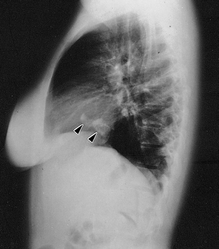 Lateral chest radiograph from a 16-year-old girl with syncope and bacterial endocarditis. The radiograph demonstrates two areas of dense calcification (arrowheads) overlying the posterior aspect of heart. The posterior-anterior (PA) view confirmed location in the heart (not shown). At surgery a calcified myxoma of the right atrium was removed. Image courtesy of Professor Peter Anderson DVM PhD and published with permission © PEIR, University of Alabama at Birmingham, Department of Pathology

| Imaging Technique | Features | Description | Advantages | Limitations |
|---|---|---|---|---|
| Two- or three-dimensional echocardiography |
|
|
|
|
| MRI |
|
|
|
|
| CT |
|
|
|
|
| Angiography |
|
|
|
|
| Chest x-ray |
|
|
|
|
References
References
- ↑ Cardiac Myxoma. Radiopedia.http://radiopaedia.org/articles/cardiac-myxoma Accessed on November 24, 2015
- ↑ Thyagarajan B, Kumar MP, Patel S, Agrawal A (January 2017). “Extracardiac manifestations of atrial myxomas”. J Saudi Heart Assoc. 29 (1): 37–43. doi:10.1016/j.jsha.2016.07.003. PMC 5247297.
- ↑ Reeder GS, Khandheria BK, Seward JB, Tajik AJ (1991). “Transesophageal echocardiography and cardiac masses”. Mayo Clin. Proc. 66 (11): 1101–9. PMID 1943240.
Editor-In-Chief: C. Michael Gibson, M.S., M.D. [1]; Maria Fernanda Villarreal, M.D. [2] Cafer Zorkun, M.D., Ph.D. [3] Ahmad Al Maradni, M.D. [4]