
Opisthotonus
For patient information, click here
Editor-In-Chief: C. Michael Gibson, M.S., M.D. [1]; Associate Editor(s)-In-Chief: Cafer Zorkun, M.D., Ph.D. [2]
Overview
Overview
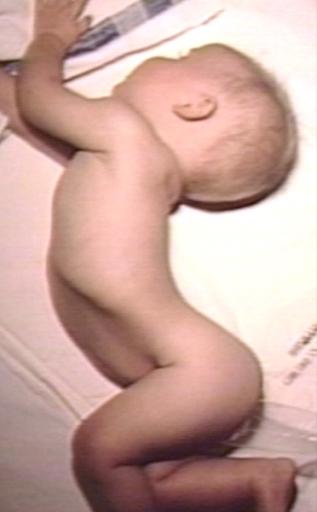
Opisthotonus or opisthotonos, from Greek roots, opistho meaning “behind” and tonos meaning “tension”, is a state of a severe hyperextension and spasticity in which an individual’s head, neck and spinal column enter into a complete “bridging” or “arching” position. This abnormal posturing is an extrapyramidal effect and is caused by spasm of the axial muscles along the spinal column. It is seen in some cases of severe cerebral palsy and traumatic brain injury or as a result of the severe muscular spasms associated with tetanus.
Pathophysiology
Pathophysiology
Opisthotonus can be produced experimentally in animals by transection of the midbrain (between superior and inferior colliculus) which results in severing all the corticoreticular fibers. Hyperextension occurs because facilitation of anterior reticulospinal tract due to removal of inhibitory corticoreticular fibers to the pons reticular formation.
Opisthotonus is more pronounced in infants. Opisthotonus in the neonate may be a symptom of meningitis or tetanus. This marked extensor tone can cause infants to “rear backwards” and stiffen out as the mother or nurse attempts to hold or feed them. Opisthotonus can be induced by any attempt at movement such as smiling, feeding, vocalization, or by seizure activity. Individuals with opisthotonus are quite challenging to position, especially in wheelchairs and car seats.
It can some times be a side effect of anti-psychotic medication or mood stabilizer, i.e. lithium intoxication.
Differential Diagnosis of Causes of Opisthotonus
Differential Diagnosis of Causes of Opisthotonus
In alphabetical order. [1] [2]
- Airway obstruction
- Meningitis
- Encephalitis
- Cerebral malaria
- Athetosis
- Cerebral hemorrhage
- Brain tumor
- Neck injury
- Drugs
- Tetanus
- Rabies
- Tay-Sachs disease
- Cerebral palsy
- Severe head injury
- Seizures
- Glutaric aciduria type 1
- Kernicterus
- Meningoencephalitis
- Strychnine
- Arnold-Chiari malformation type 3
- Hereditary methemoglobinemia, recessive, type II
- Perinatal hypophosphatasia
- Phenothiazine, Perphenazine
- Antenatal infection
- Schinzel-Giedion syndrome
Physical examination findings
Physical examination findings
-
 Opisthotonus: Meningitis, One Year Old Child: Pus in Subarachnoid Space causes Neck Muscle Spasms
 -
 Opisthotonus: Perinatal Hypoxia; Note Spastic Quadriparesis,


External links
External links
Looking for the patient version?
© 2026 MyEClinic – IFTM Institut für Telematik in der Medizin GmbH